VSI Simulator Production Process
Take a look at this slide show to see the process behind our simulators!

This photo shows the fiberglass horse with the access hatch cut into place. This will allow the instructors to place a real digestive tract into the unit to facilitate palpation. finished units will have a urethane tub to contain the organs, as well as a reproduction pelvis. The bottom of the belly also has a replaceable plug that simulates the abdominal wall of the horse and can be cut and punctured with a canula to simulate the belly tap procedure.


One of the finished prototype units being used at the University of Calgary Veterinary School. It has a simulated rectum, anus, and vagina, as well as a reproduction pelvis. The unit also has a belly tap function and provisions for using real equine digestive tracts.


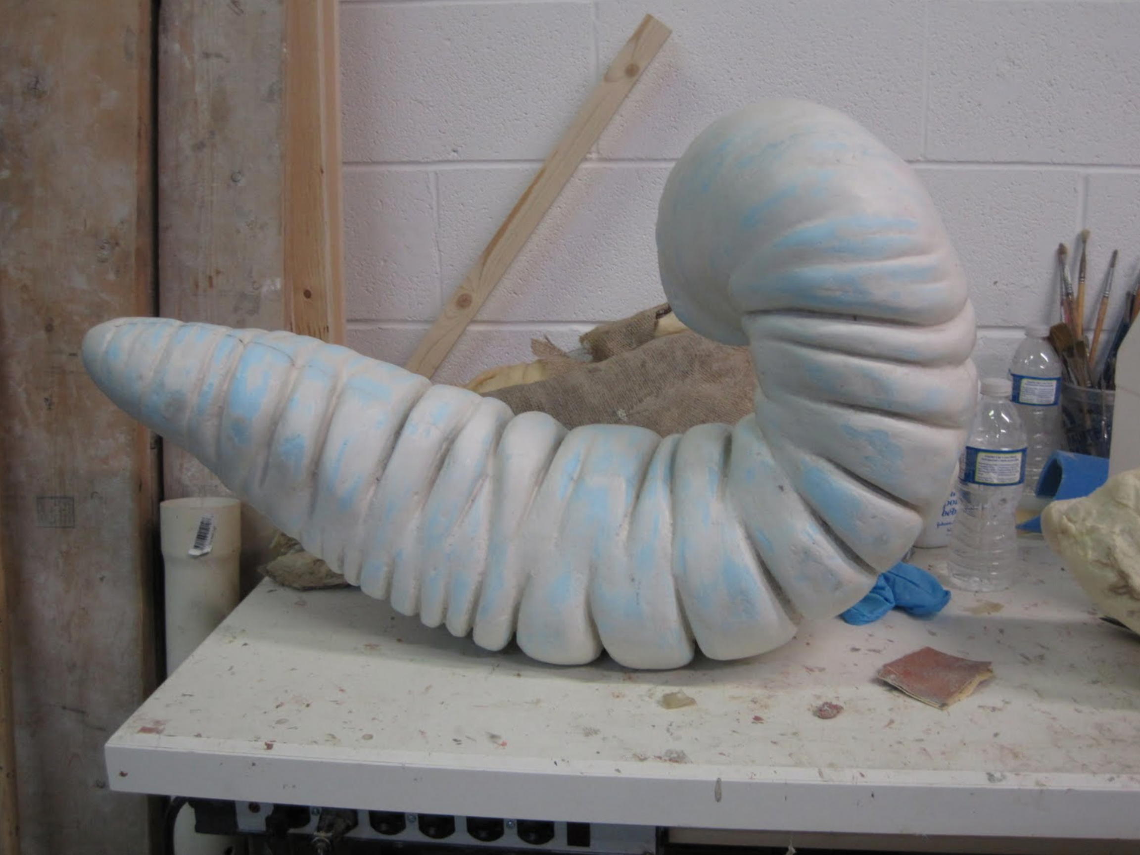
The cores of the equine colon with the biological materials removed. These will have any flaws repaired and be used in the next stage of creating an equine colic simulator. Various materials will be tested in order to achieve a realistic feel and durability.

A portion of the 23 pieces of fiberglass jacket that will support the rubber portion of the Holstein mold. Highly detailed copies of the original sculpture can now be produced in fiberglass. These will be the basis for the Holstein dystocia simulator.


This photo shows the core of a portion the equine large intestine. It will be used to make a reproduction of the colon to simulate equine colic. the entire equine colon will be reproduced including the secum and a portion of the small intestine.


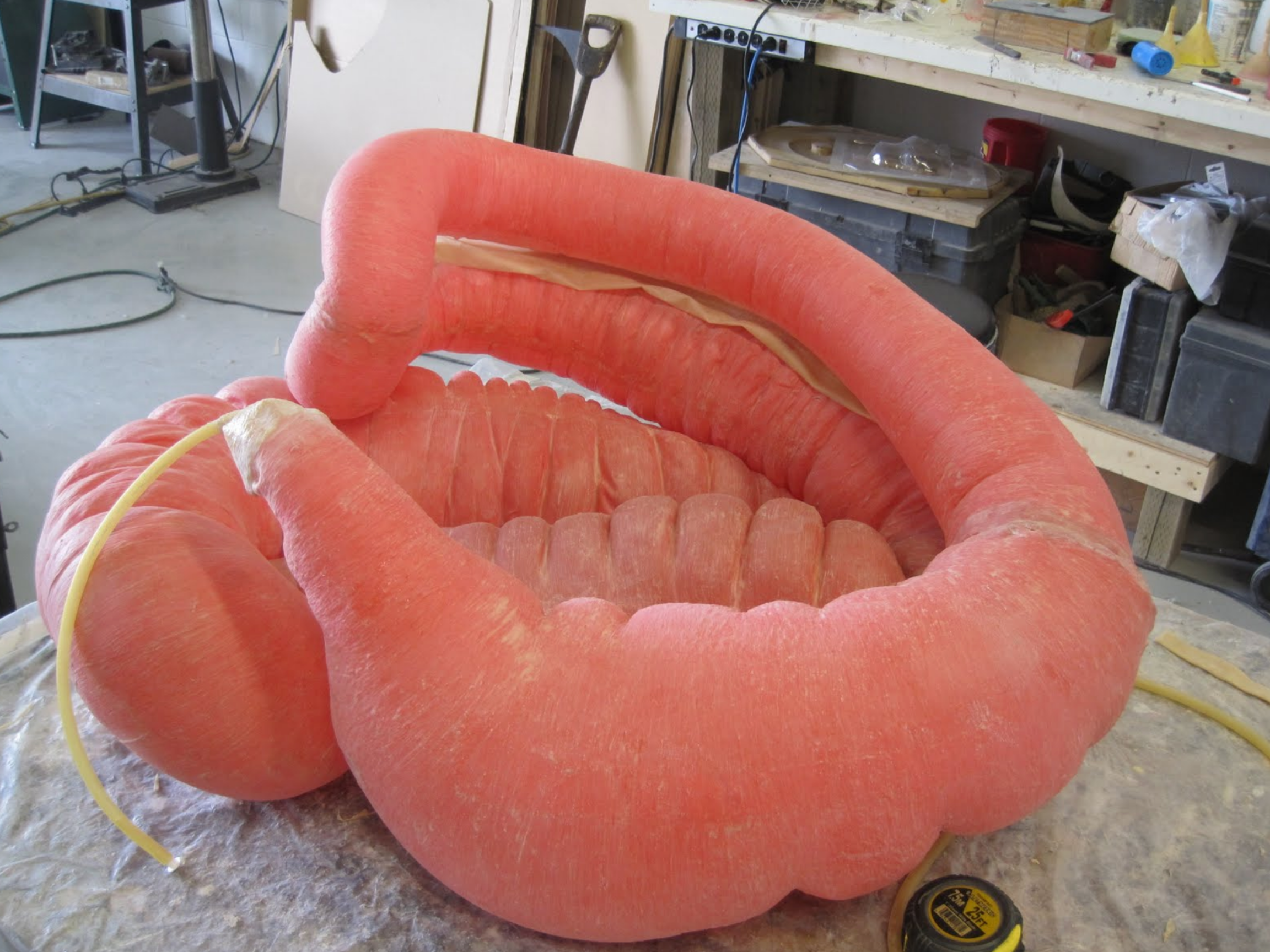
This photo shows a portion of the equine colon test piece being inflated to evaluate the feel of the chosen material. Once we are pleased with the result, veterinary professors from the University of Calgary Faculty of Veterinary medicine will further evaluate the simulator.


The nearly complete equine colon model. A portion of the small intestine and small colon will be added as well. The finished units will have chambers that can be inflated separately to simulate various equine colic issues. These units will mount into the existing equine simulators already in use at the University of Calgary Faculty of Veterinary Medicine.


Work now continues on the equine colic simulator. Here the various sections of the equine large intestine are being prepared for assembly. Once assembled they will be fitted into the fiberglass equine model along with representations of the kidney’s and spleen. These sections can be inflated to provide a realistic feel for palpation exercises.


This photo shows the inflated equine GI tract model, ready for final paint and detailing. The finished piece will then be mounted into the horse model along with representations of the spleen and kidney.

The fiberglass model with head and legs attached permanently for durability. the model will be fitted with the colon model, as well as spleen, kidney and nefrosplenic ligament.
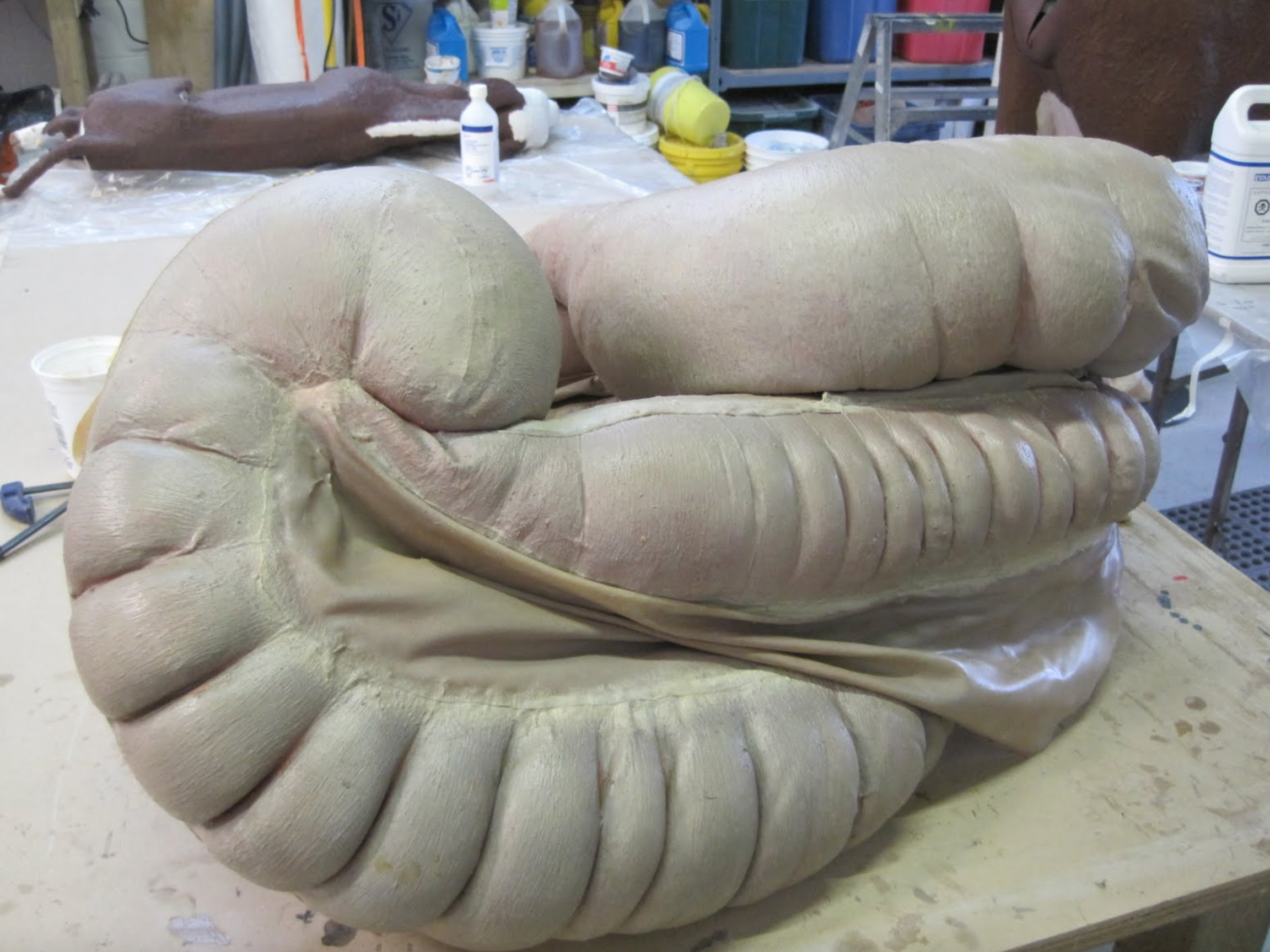
This photo shows the finished prototype equine GI tract. It can be positioned to simulate various colic conditions. Two more units will be made to simulate an impaction of the cecum and a pelvic flexure impaction.
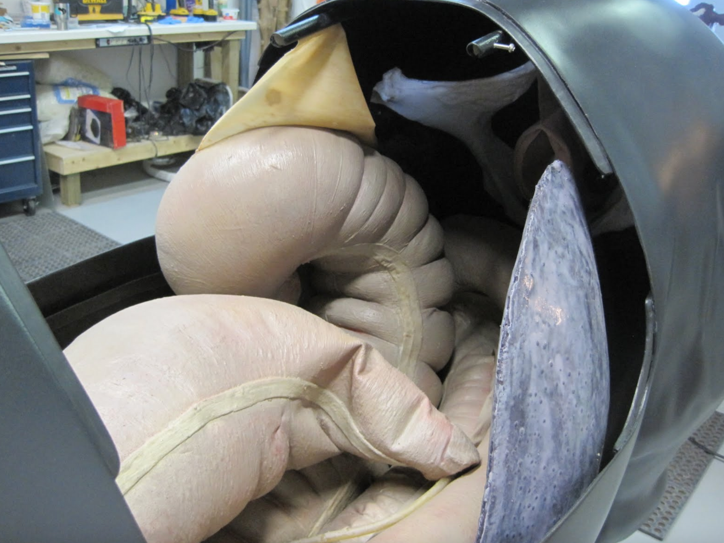
The equine GI tract positioned in the model, along with the representation of the spleen. There will also be a model of the left kidney and renosplenic ligament for palpation training.
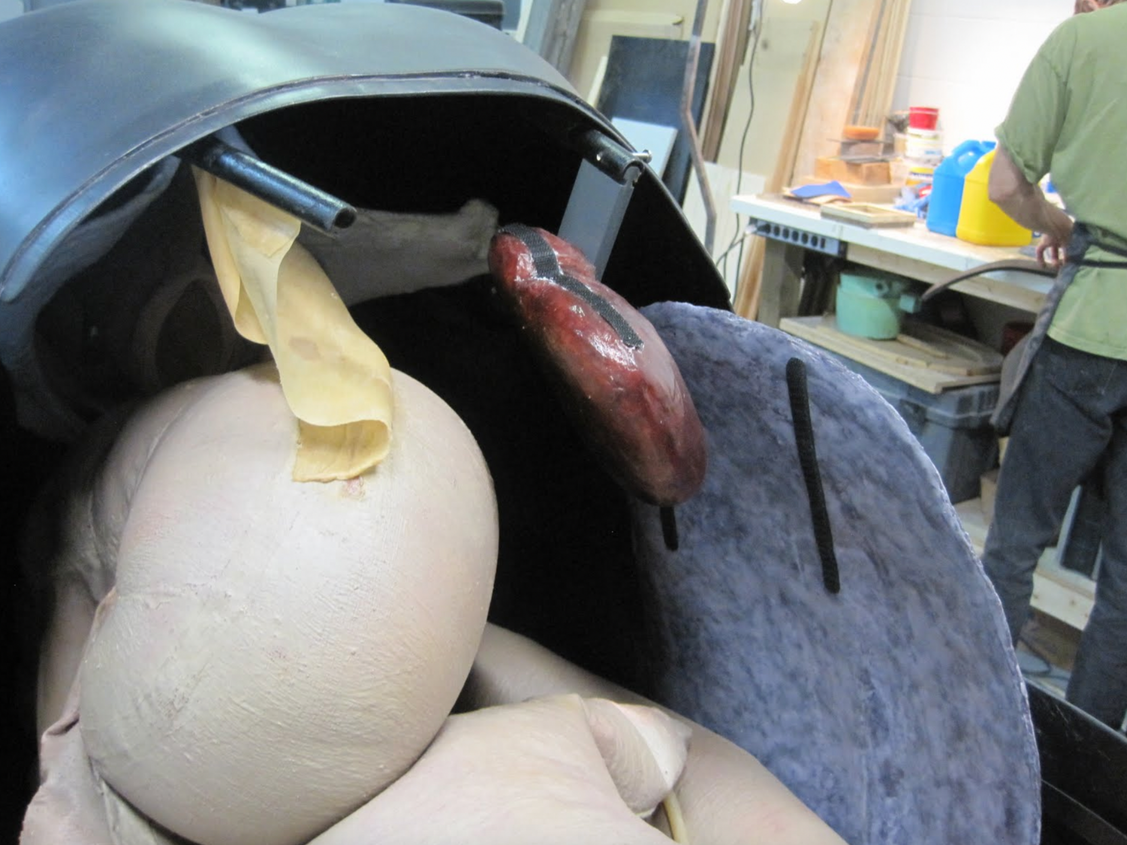
Here the left kidney is in place along with mounting positions for the renosplenic ligament. Being that this unit is a prototype, all aspects will be refined once testing is completed.


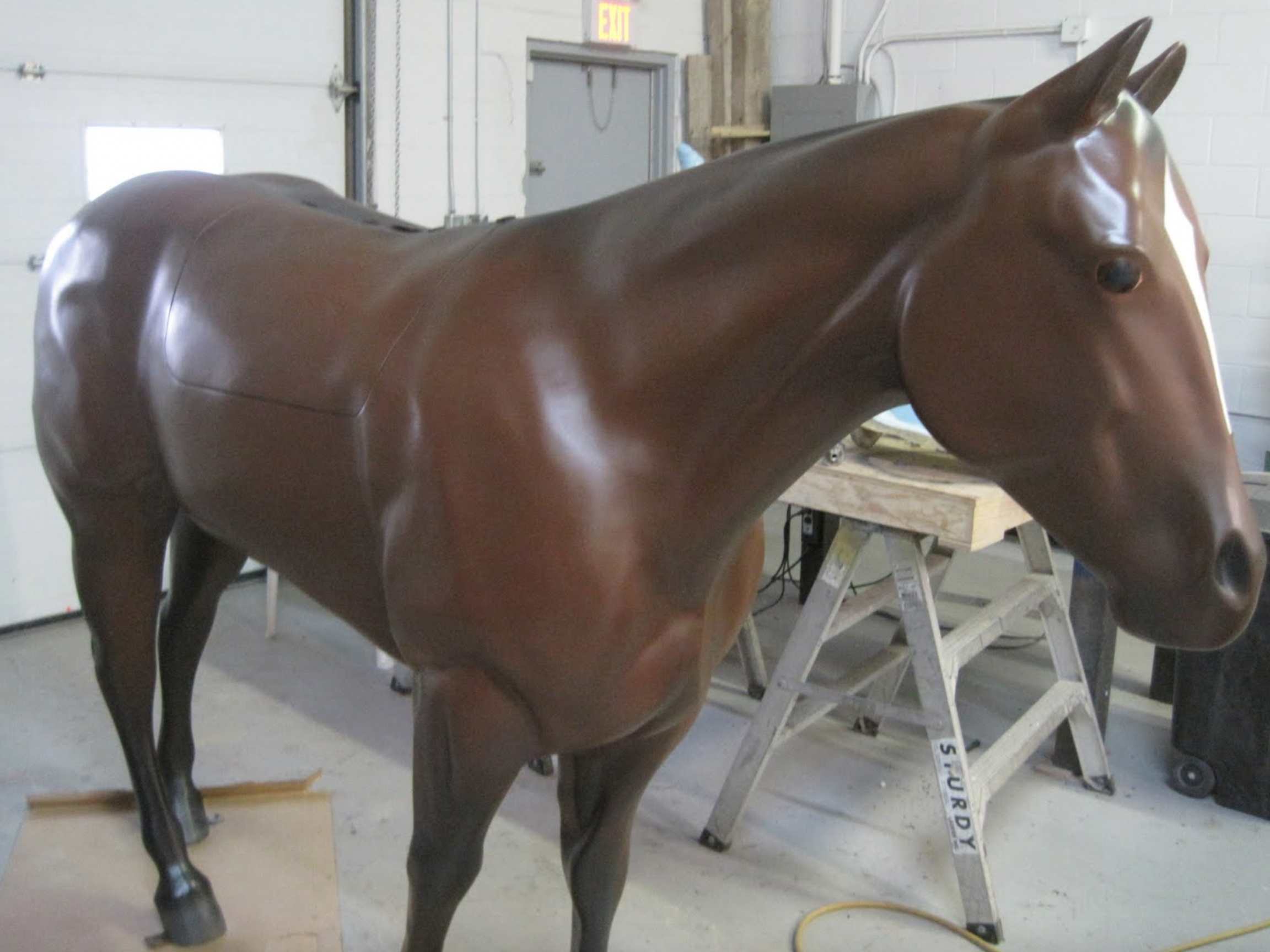
The third horse that has been fitted with a GI tract simulator, and a belly tap function. The unit will also have a spleen, reno–splenic ligament and kidney models.
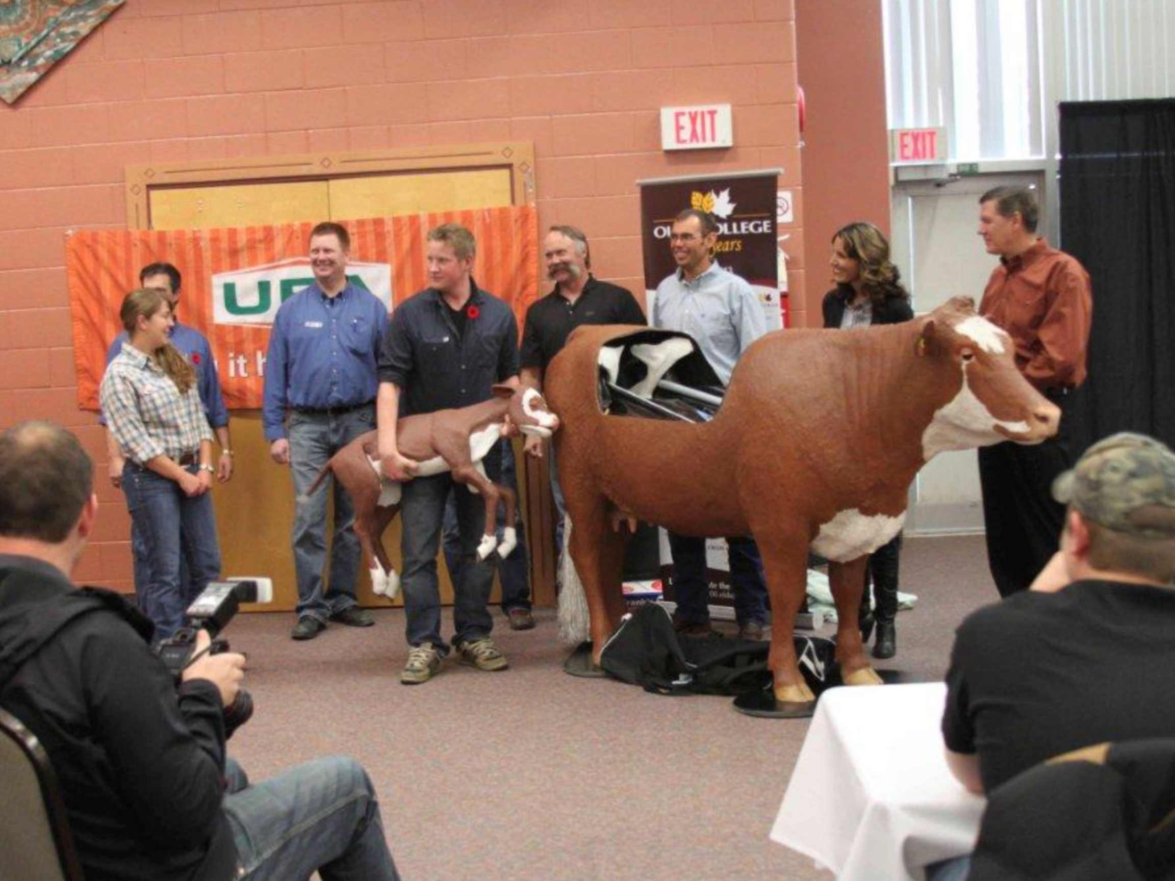
“Lucy” and “Lou” being presented to Olds College staff and faculty by the UFA team. The bovine dystocia simulator was purchased and donated to Olds Agricultural College by UFA which has a long-standing association with the college.
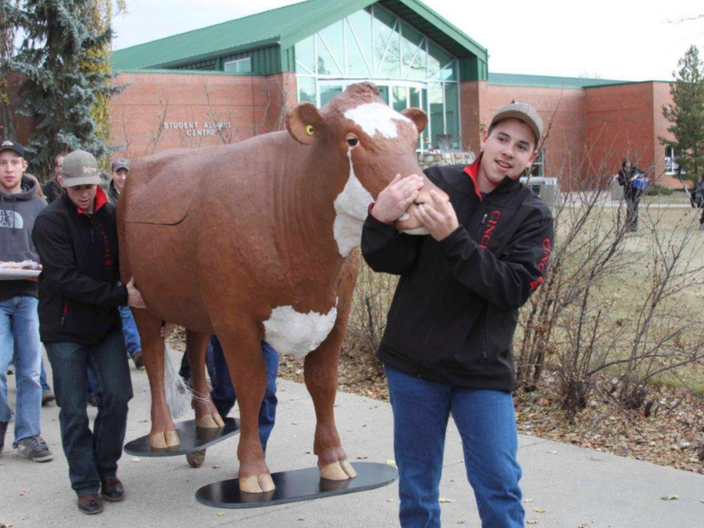

“Lucy” in the classroom. Students can now learn various dystocia issues and procedures in the classroom and practice those solutions without causing discomfort or endangering a live animal or themselves. This allows them to build confidence before moving on to producers live animal.
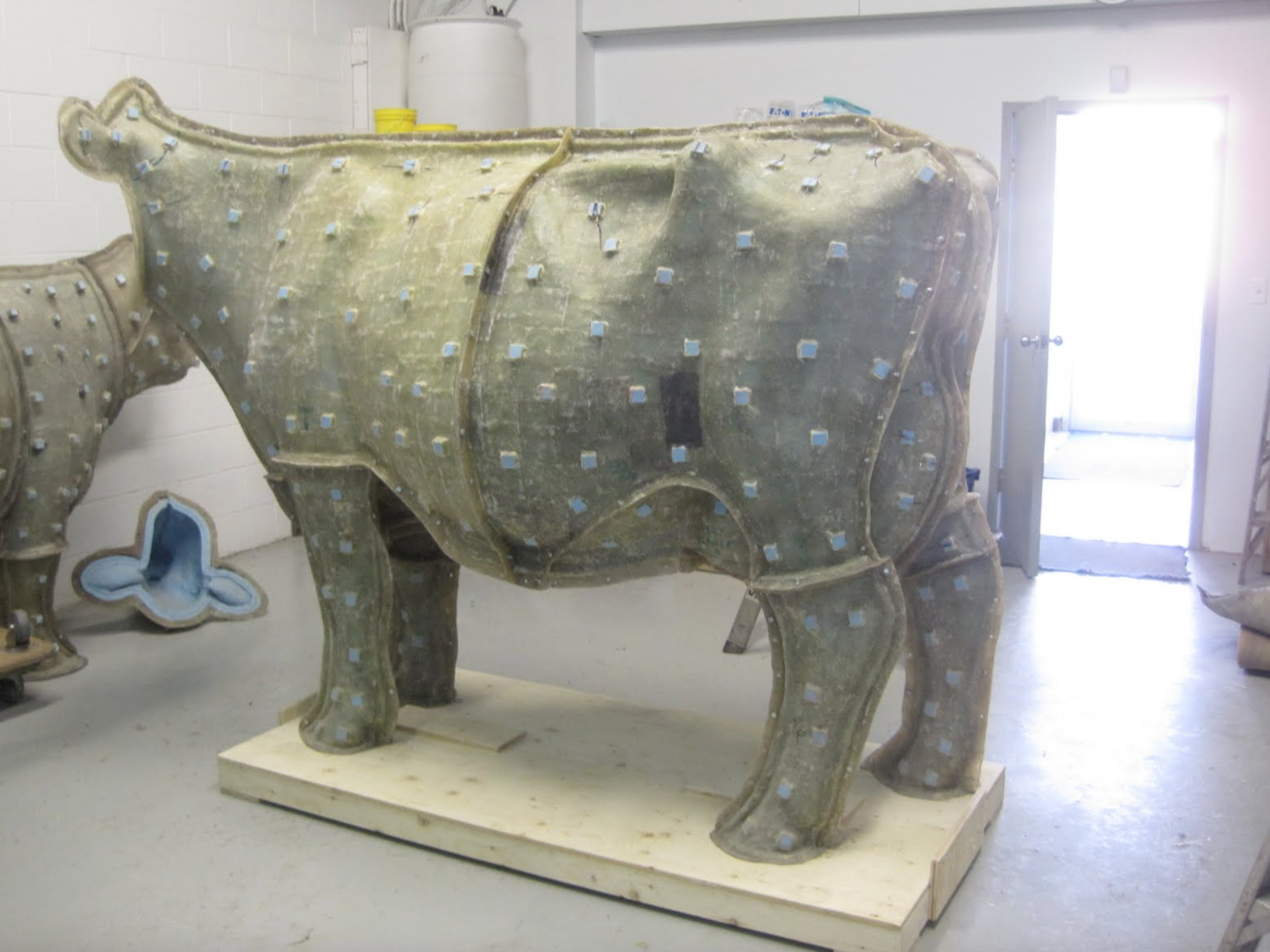
Our true type Holstein mold is being readied for the casting process. We are now using epoxy resins, instead of the more conventional polyester resins, due to their low VOC’s, higher strength and flexibility, and greater chemical resistance.
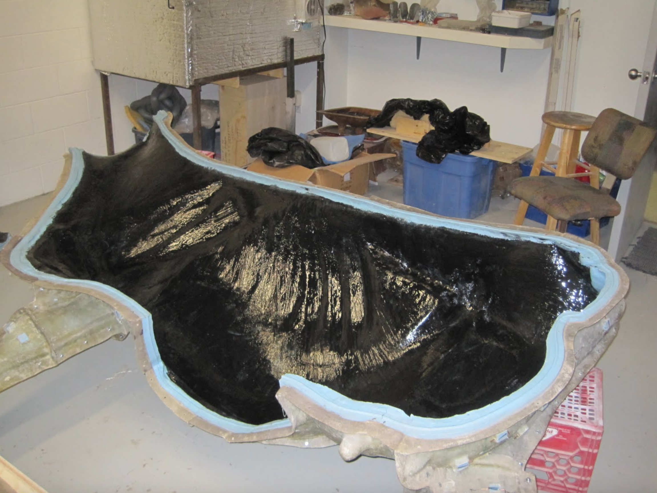
One side of the Holstein mold, with the first layer of epoxy resin applied. Several more layers of fiberglass will be applied, infused with epoxy resin. The two halves will then be joined together to complete the body.
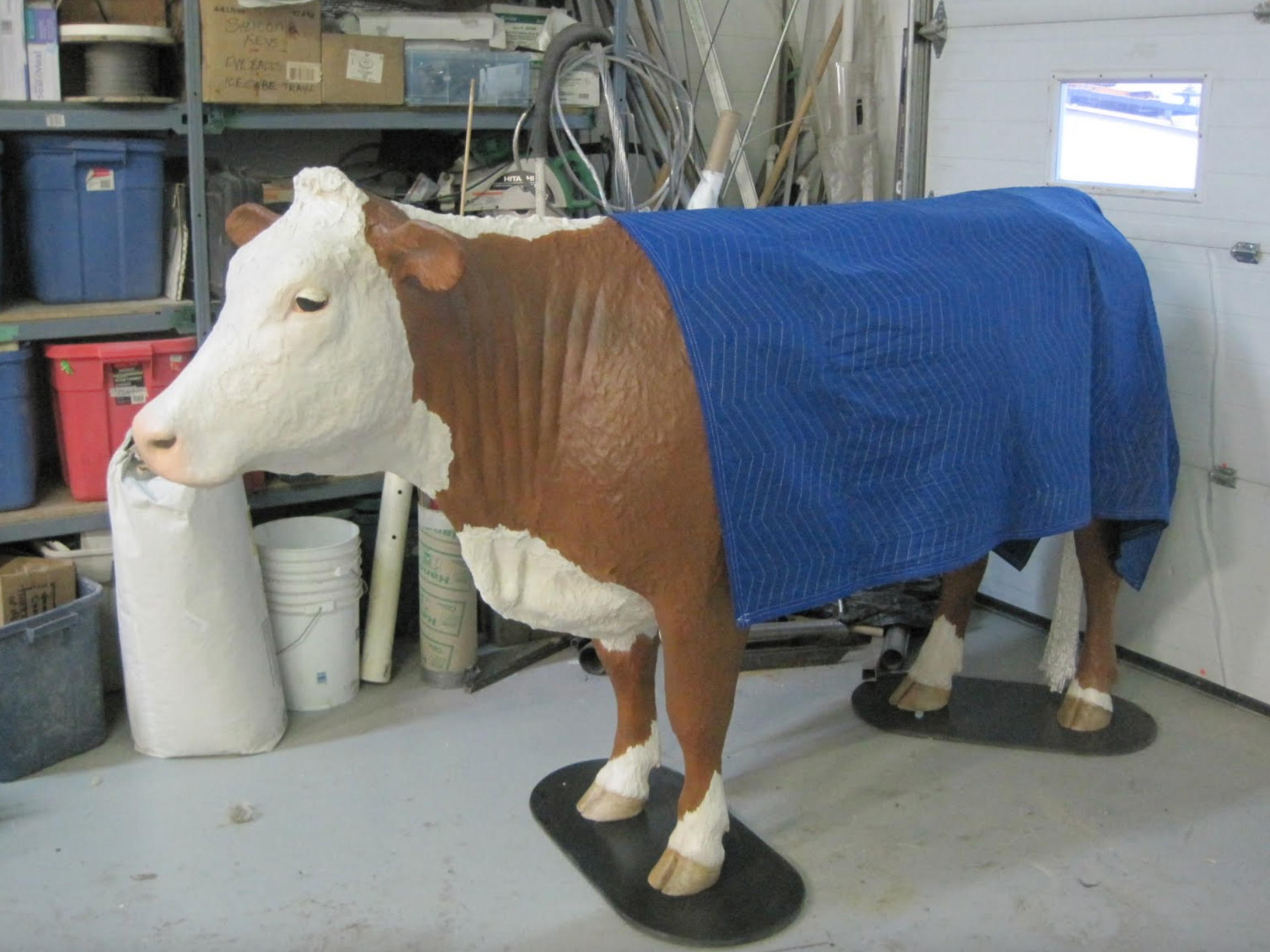
This completed Hereford model traveled to the University of Saskatchewan Western College of Veterinary Medicine for a demonstration of the simulator for members of the faculty. We also demonstrated an equine colic simulator prototype and gathered feedback on both units.
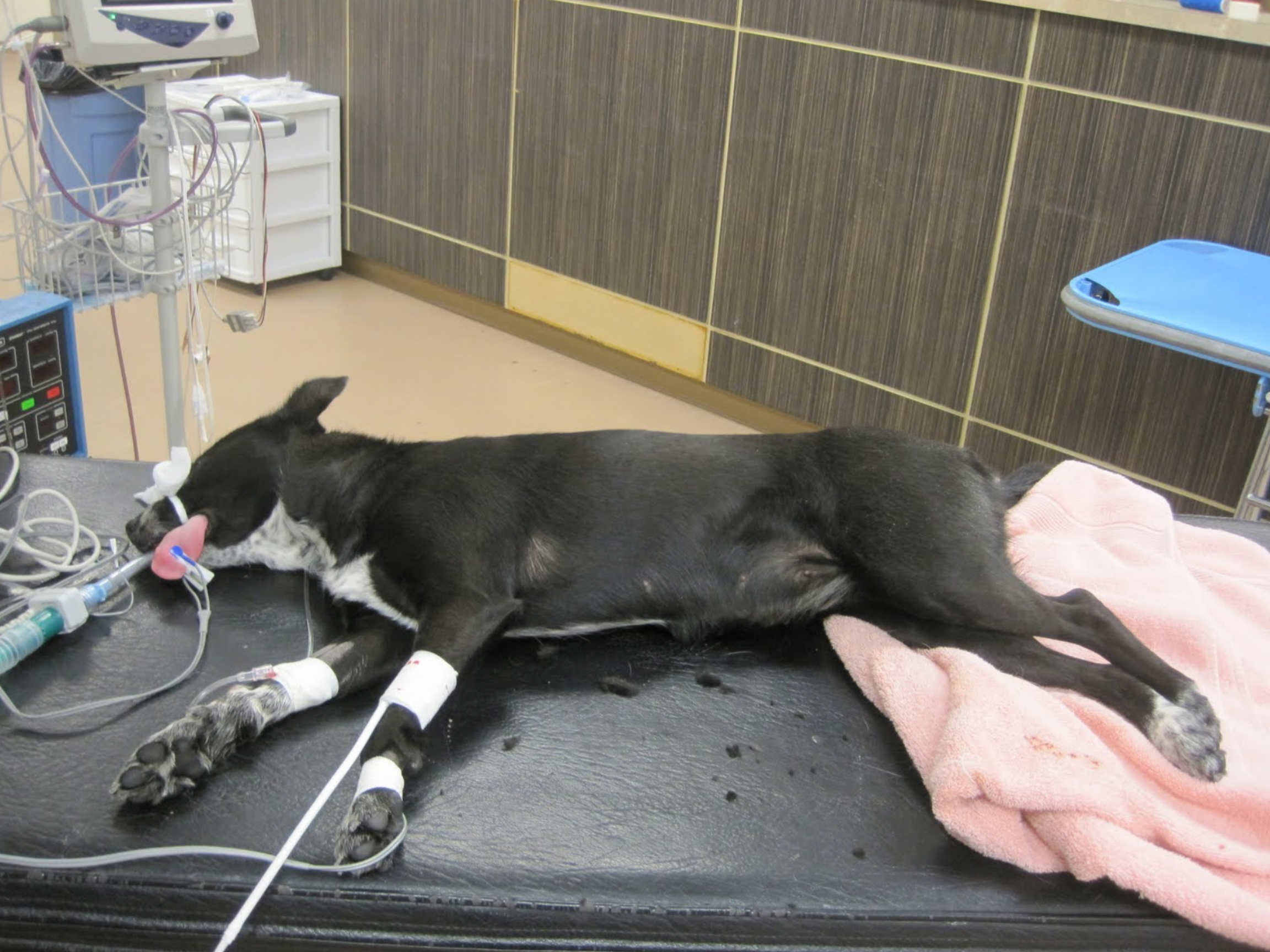
We recently participated in a canine spay procedure by Dr. Audrey Remedios in order to better understand the structures involved and how they feel. This will allow us to better recreate these structures in a spay simulator. We were also shown how an esophogastric tube would be inserted for a better understanding of that procedure as well.


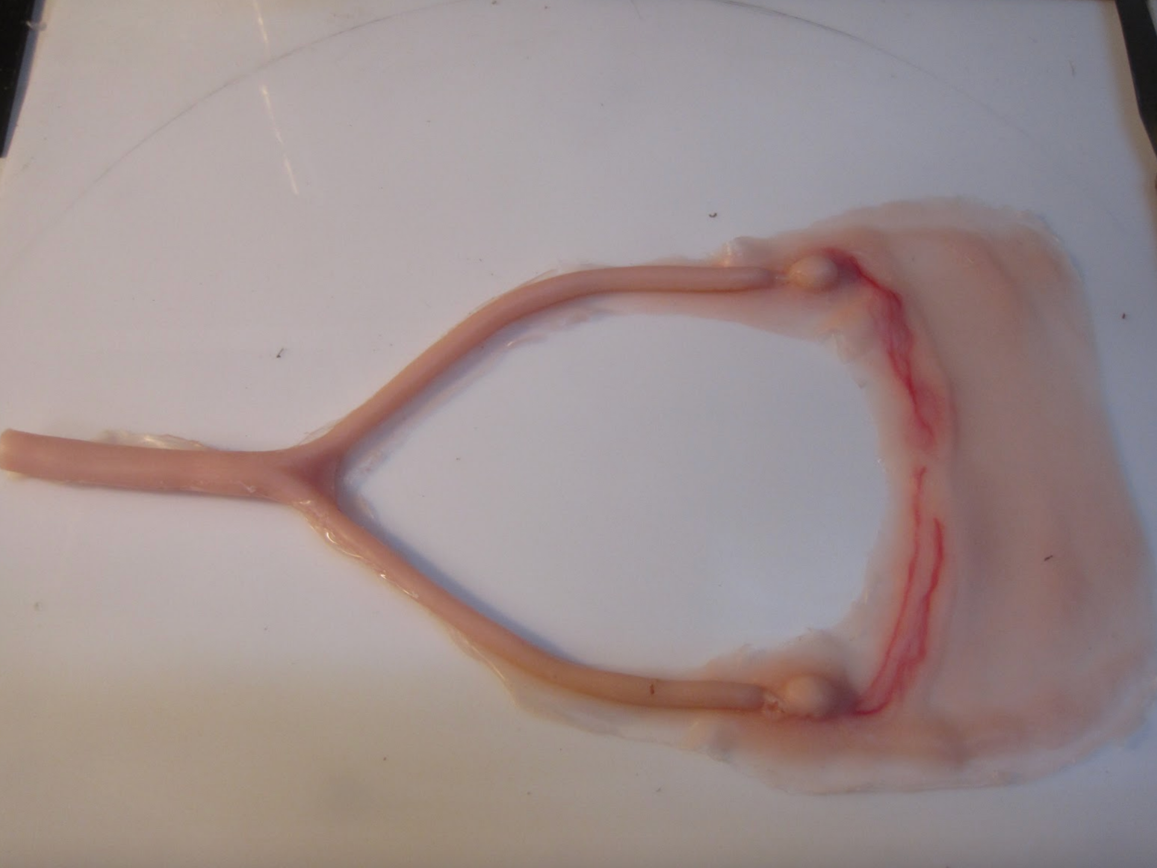
Here we have a prototype canine uterus model. It will be fit into the base unit and be anchored appropriately for a realistic spay procedure. This will be the consumable portion of the spay model, along with a suturable skin, similar to our exiting suture pad .
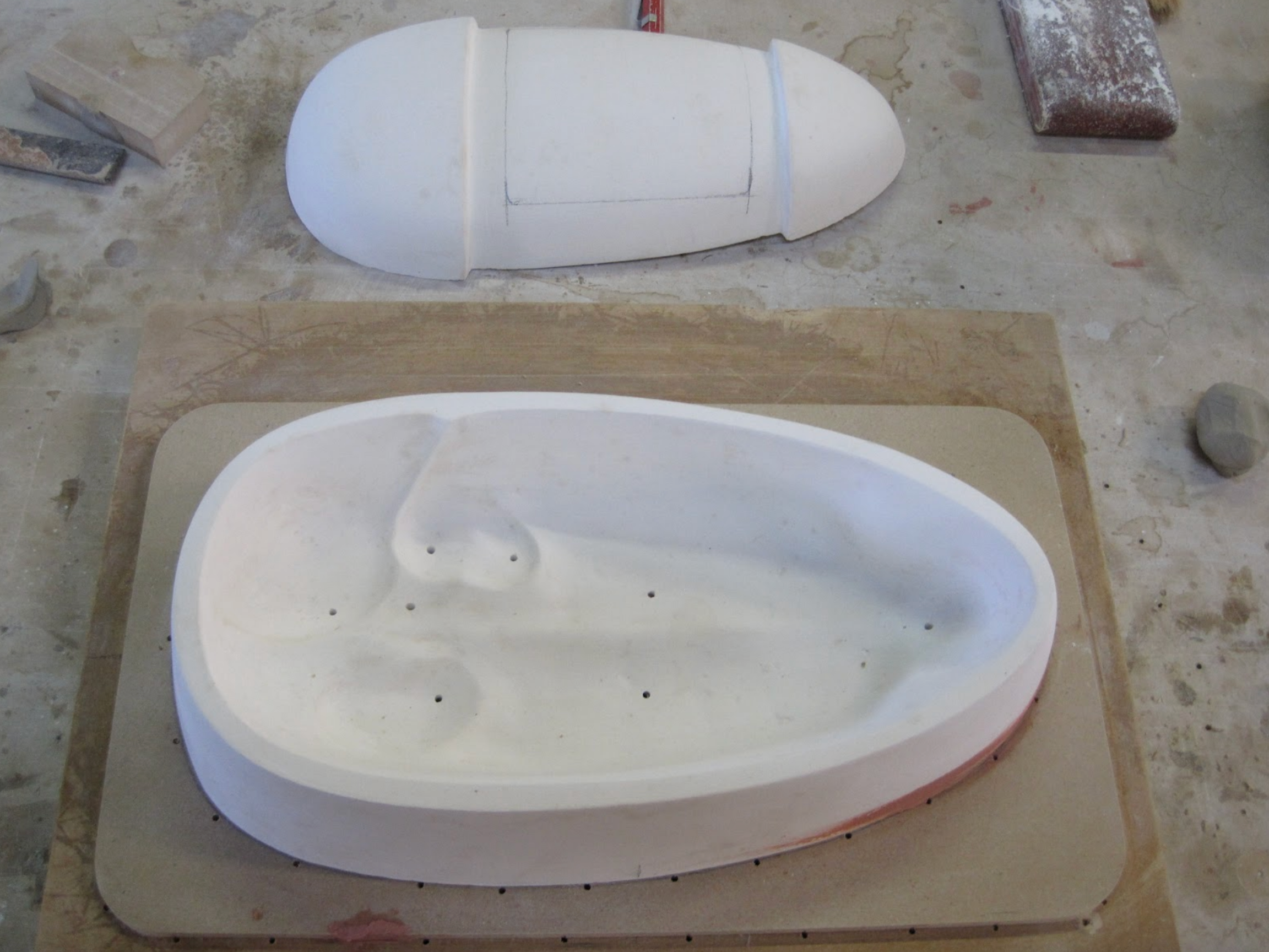
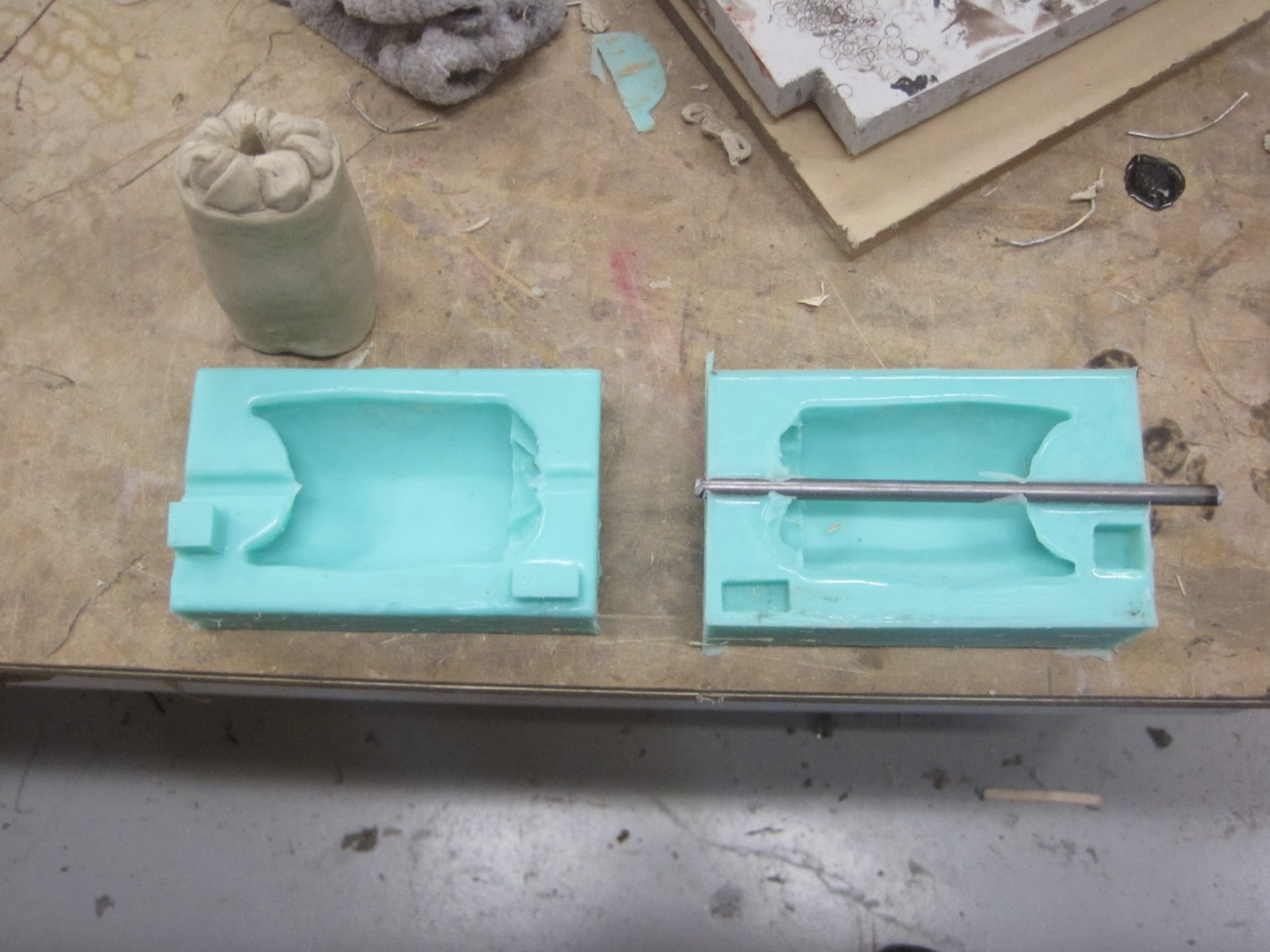
In this photo we have the two positive forms for the prototype canine spay unit. The prototypes will be a thermo-formed shell which will house all the soft internal components.
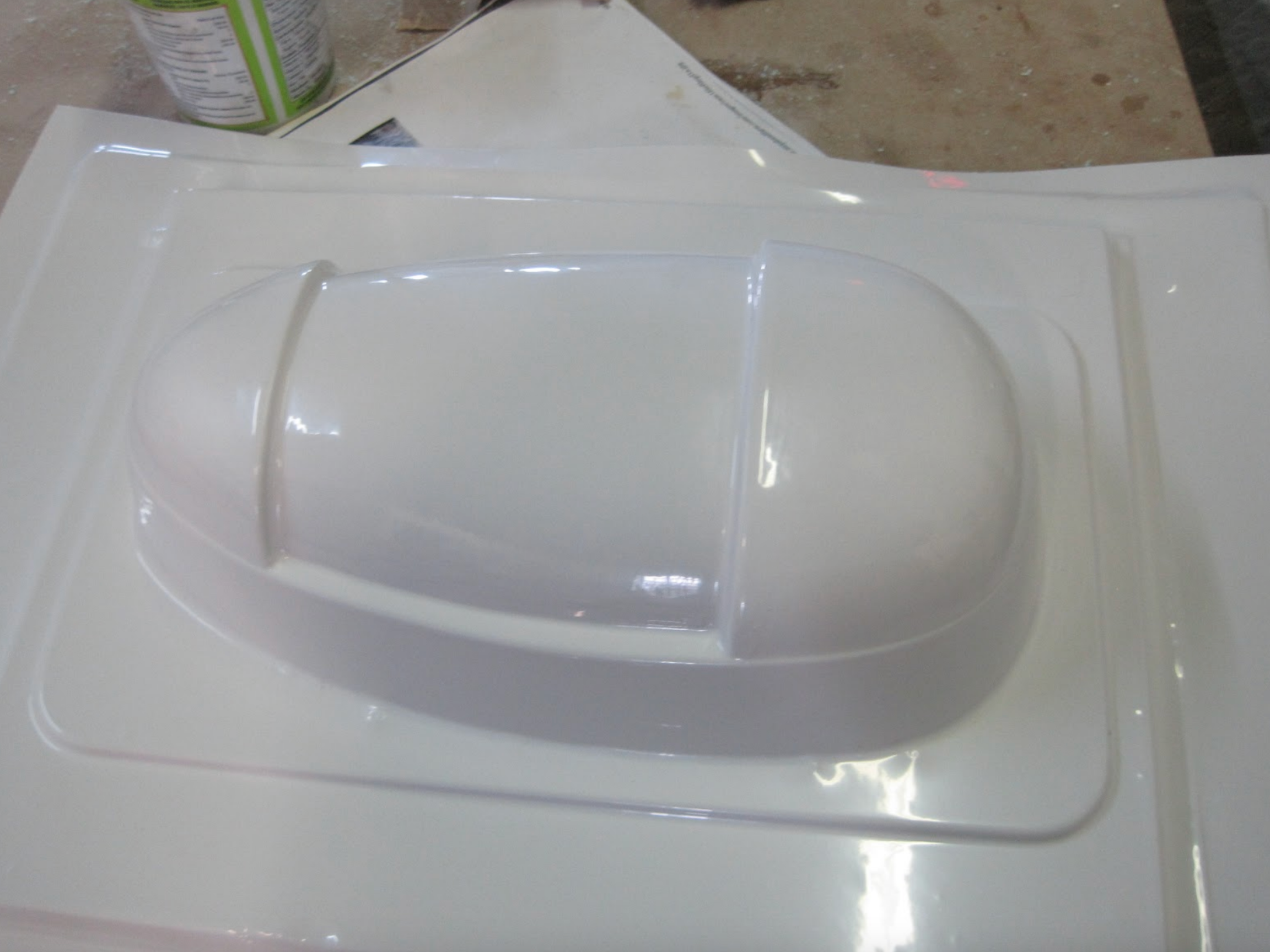
The first copy of the prototype’s outer shell. Once trimmed it will be fitted with suction cups and the inner base unit, that contains the uterus, kidneys, spleen and intestines. The outer shell will accept a suture pad for making the incision and suturing it closed.
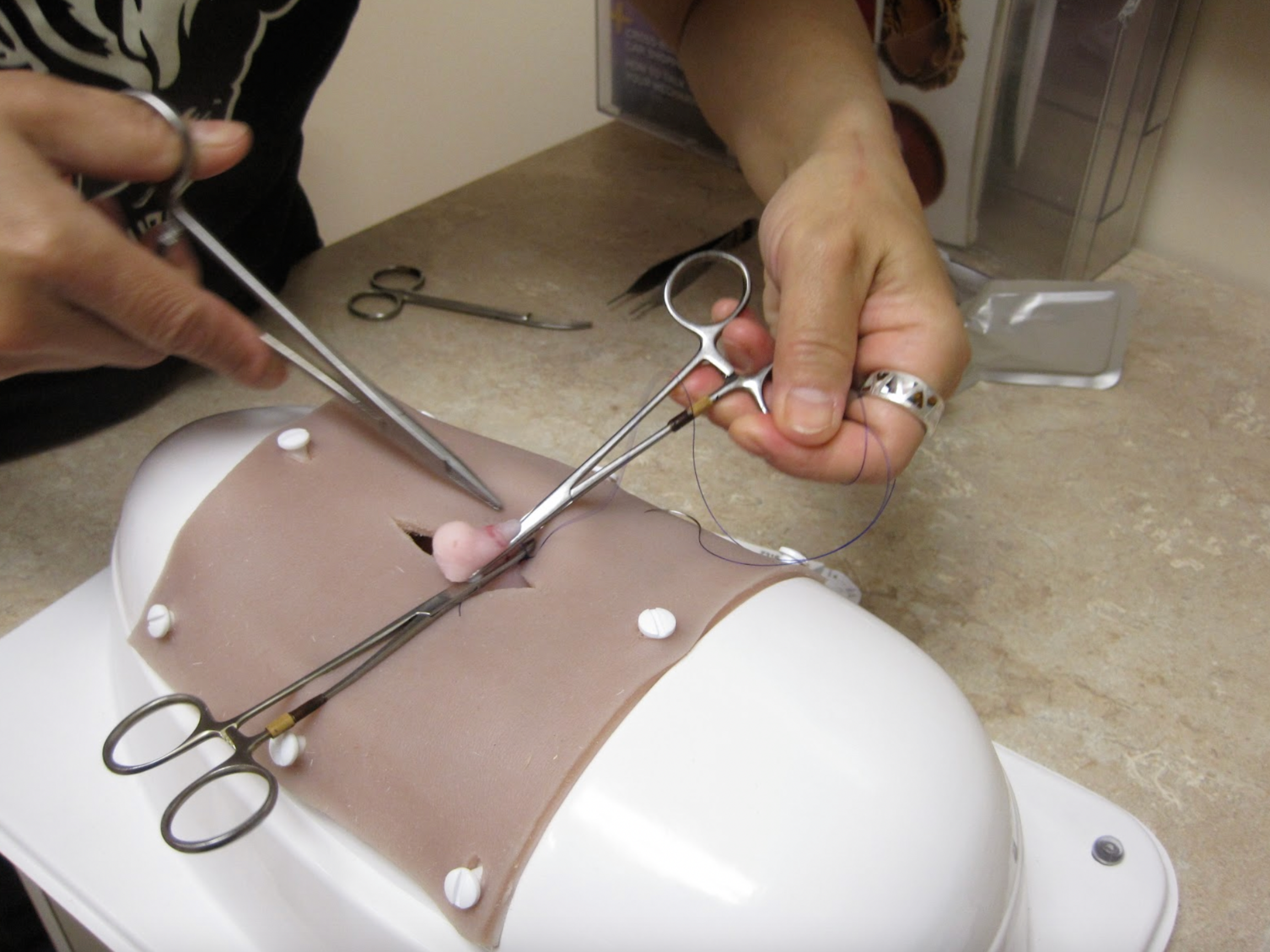
Here we see Dr. Audrey Remedios performing a simulated spay on a prototype unit to evaluate its performance and realism. Through this trial we determined what needed to be changed in order to make the spay simulator work more realistically. The only consumable parts of this unit are the uterus and skin. The skin is sized to work with our suture pad bases as well, so it can be cut and sutured several more times after the spay procedure if desired. With this unit we have endeavored to keep the procedure costs as low as possible.
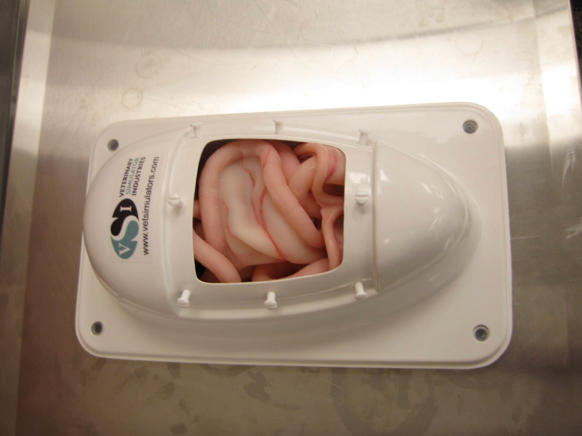
This photo shows the completed prototype canine spay unit. Ten units have been built in order to test them in a classroom situation. Hopefully this will identify any possible problems or shortcomings with materials or design. This design uses a suture training pad for the skin incision.
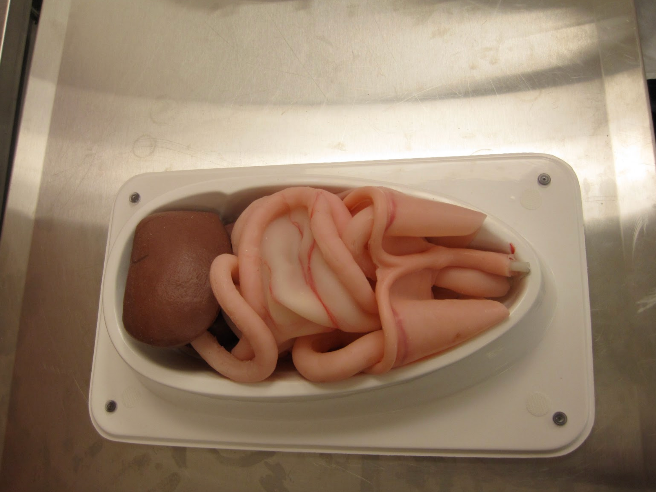
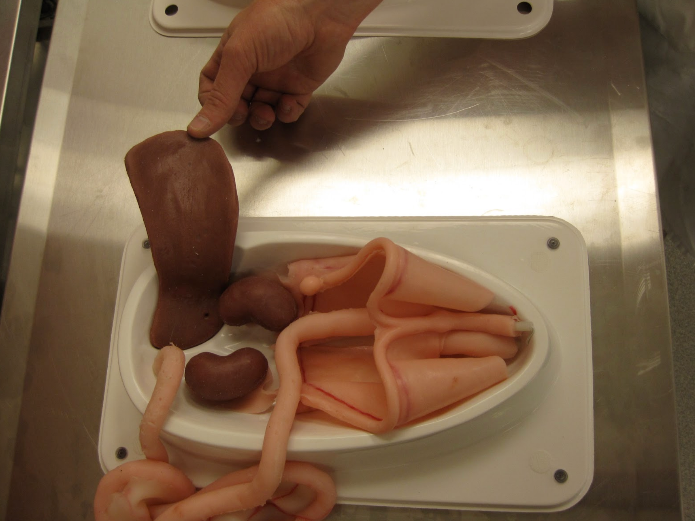
Spleen pulled back and intestines removed showing the kidneys and uterus. The uterus along with the broad ligament, suspensory ligaments and skin can be quickly and easily replaced to re-set the spay unit.
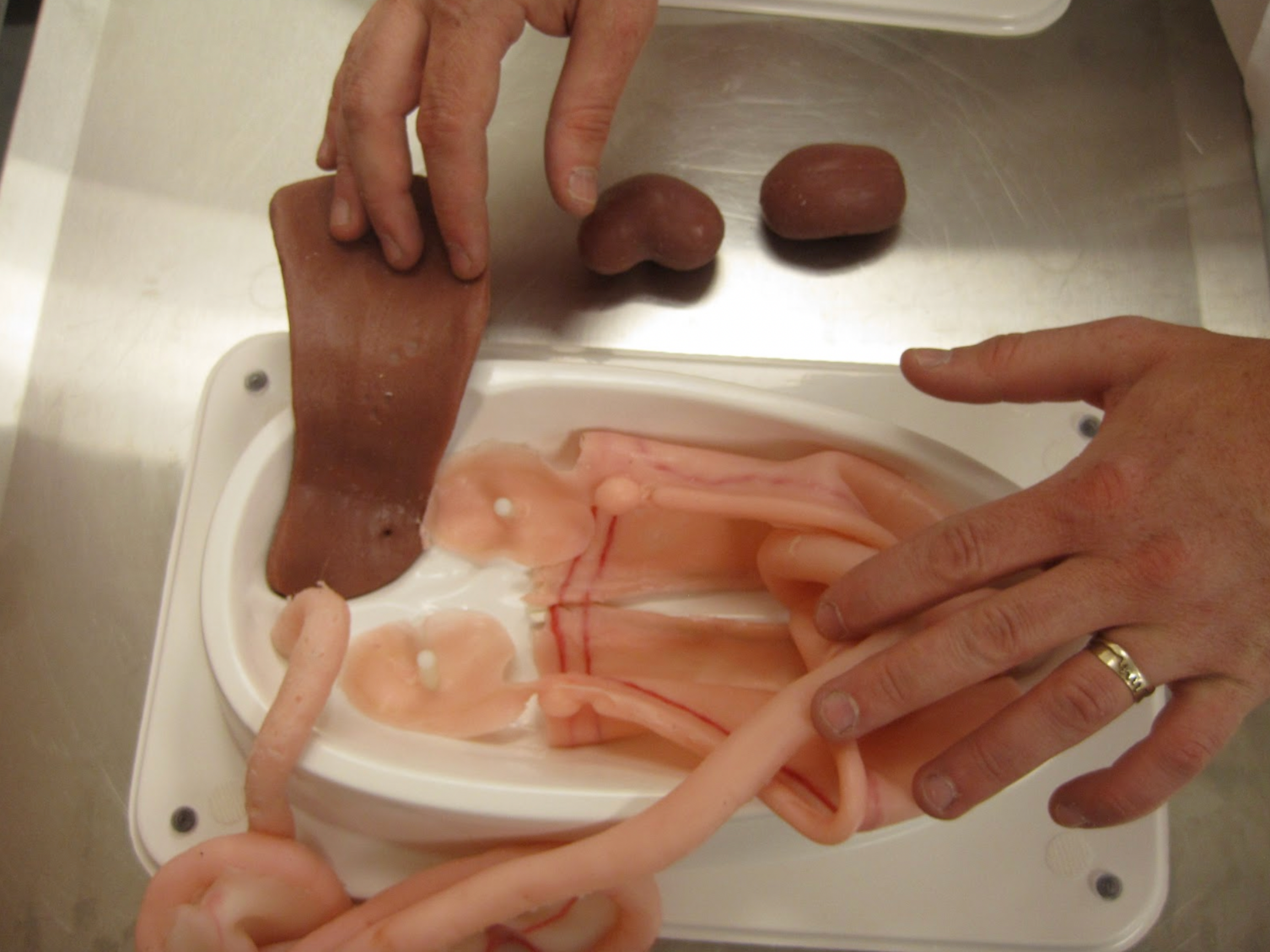

This photo shows an equine left kidney. The University of Calgary’s Veterinary anatomy department provided us with samples of the equine left kidney, spleen and reproductive tract along with a sample the bovine reproductive tract. We will be creating replica’s of all these structures for both our equine and bovine simulators.

This equine reproductive tract will be the basis for the sculpture we will create in order to reproduce it. The actual structure cannot be effectively molded due to its softness. We will create a sculpture, mold and cast it in a extremely soft material. The finished reproductive tracts will be suspended in the equine simulators to allow for palpation training.

We have begun the process of creating the equine leg injection model. We have positioned the leg in preparation for the molding process. Here we see the equine hind leg ready for the coating of alginate.

In this photo an equine front limb has been positioned in preparation for molding.

Here the leg has been removed from the alginate. The mold has been reassembled and will be filled with plaster to create a positive reproduction of the leg.

The plaster cast will now be cleaned up and any imperfections repaired before the final molding process will begin. The biological legs will be returned to the UCVM anatomy department where the flesh will be removed and the skeleton cleaned. Then the process of molding the bones of the leg will start.

The plaster cast has captured all the detail of the hoof, but still requires a small amount of clean-up.
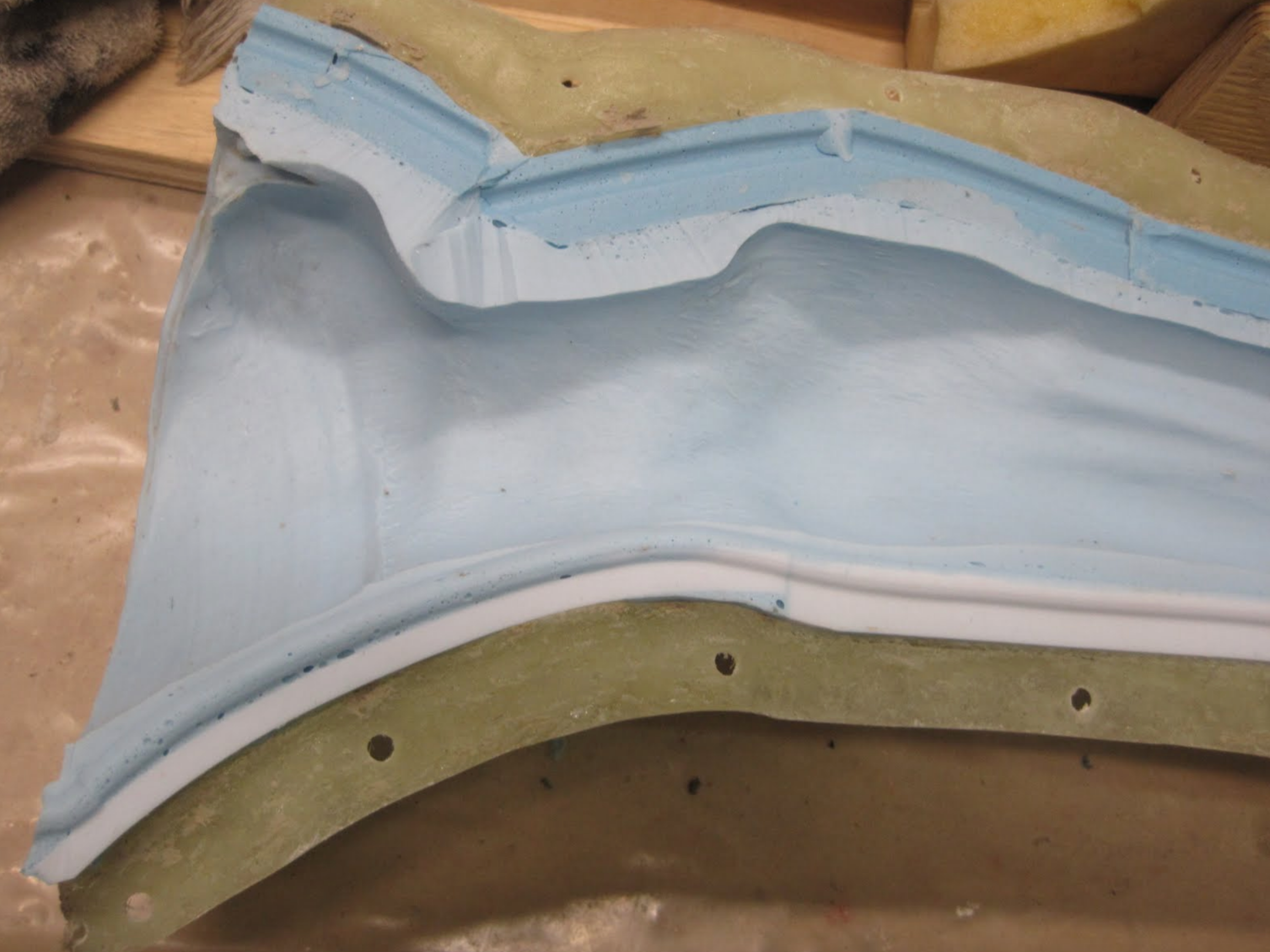
This photo shows the interior of the finished mold, along with the fiberglass jacket.

The plaster cast has been repaired and has had several coats of silicone molding material applied.
Once molds of the exterior of the leg have been completed we will begin molding the skeletal structures. Once cast, the bones will then be articulated and the reproductions of the muscles, tendons, nerves and synovial membranes, can be sculpted on the skeletal structures.

The equine leg mold now has the keys attached and is ready to have the bottom of the hoof poured. It then will have a fiberglass jacket applied, in three sections. then the mold of the leg exterior will be complete.

Here we see the equine leg bones ( cannon and splint ) with the clay dam and mold box. Silicone will now be poured into the box to create the mold. All the bones of the lower foreleg will be reproduced this way. Once all are cast in a hard urethane they will be articulated and fit into the exterior leg mold.

One half of the equine fore limb bone mold has been poured. This half will be coated with a release agent then the other half can be poured. This procedure will be repeated for all the bones of the fore limb.
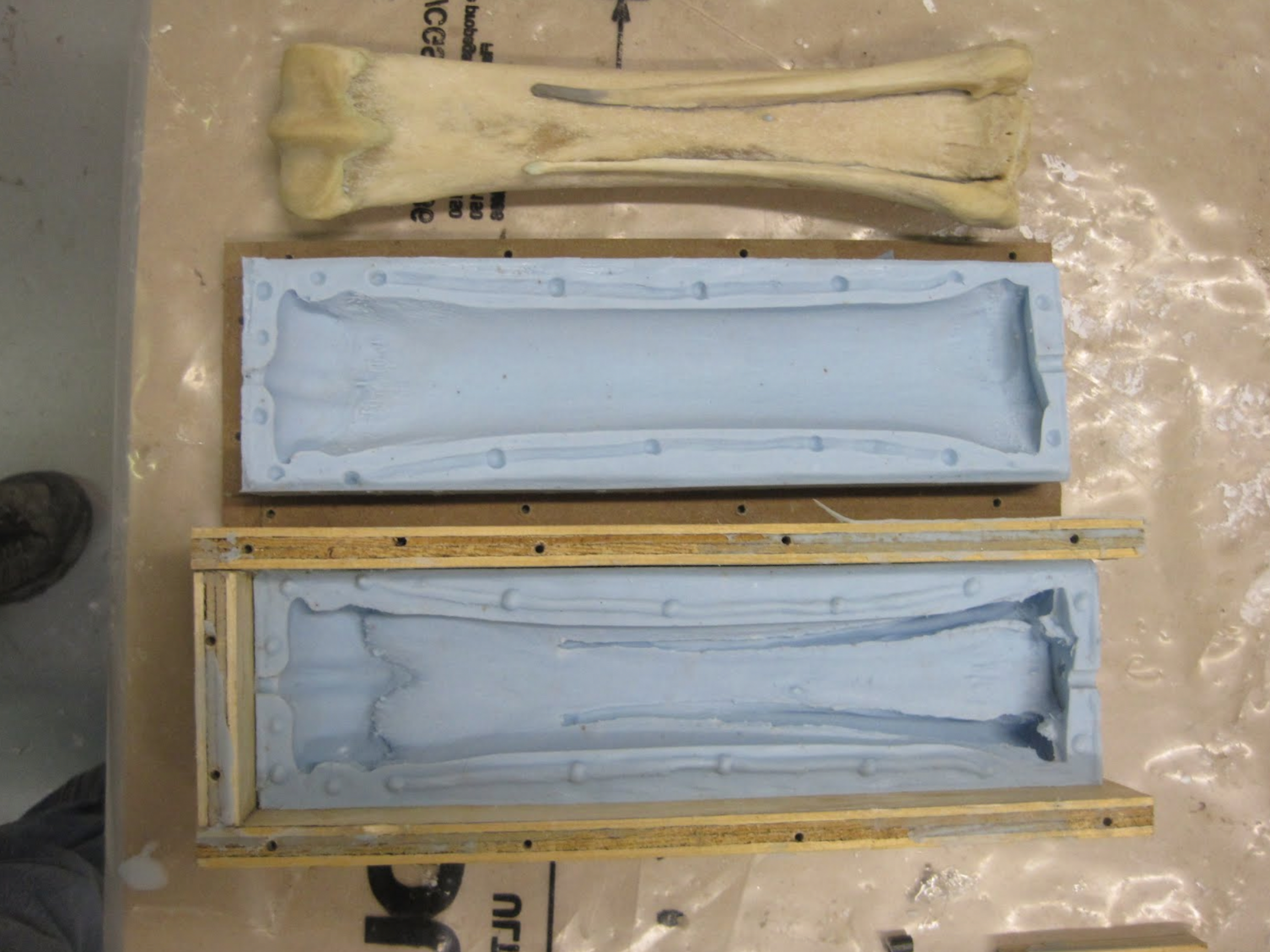
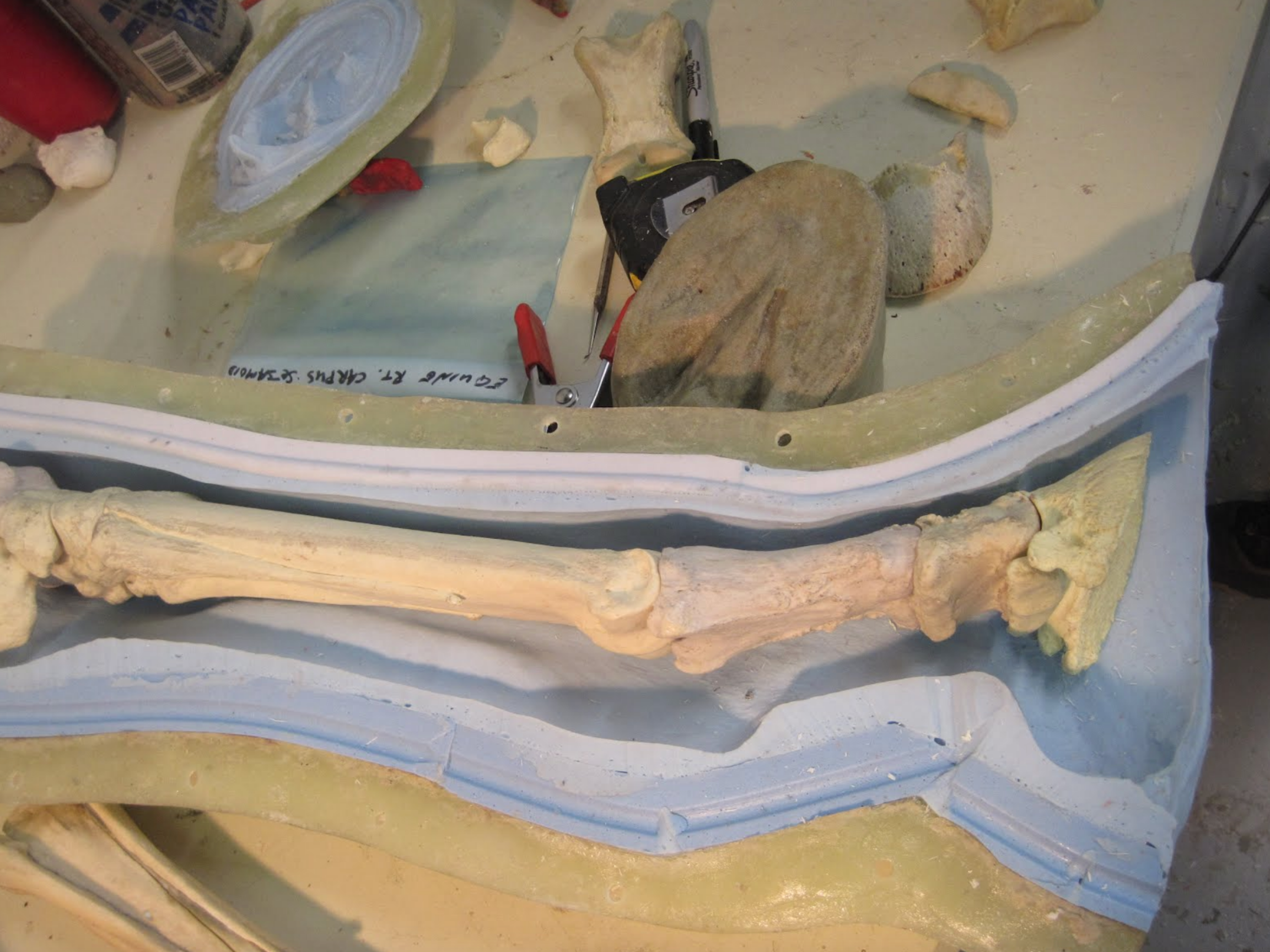
The leg bones have now been cast in a urethane plastic. They have been assembled and attached together to allow the skeletal structure to be registered within the exterior leg mold. This will allow us to begin work on the structures between the skin and bones( muscles, tendons,and synovial pouches).
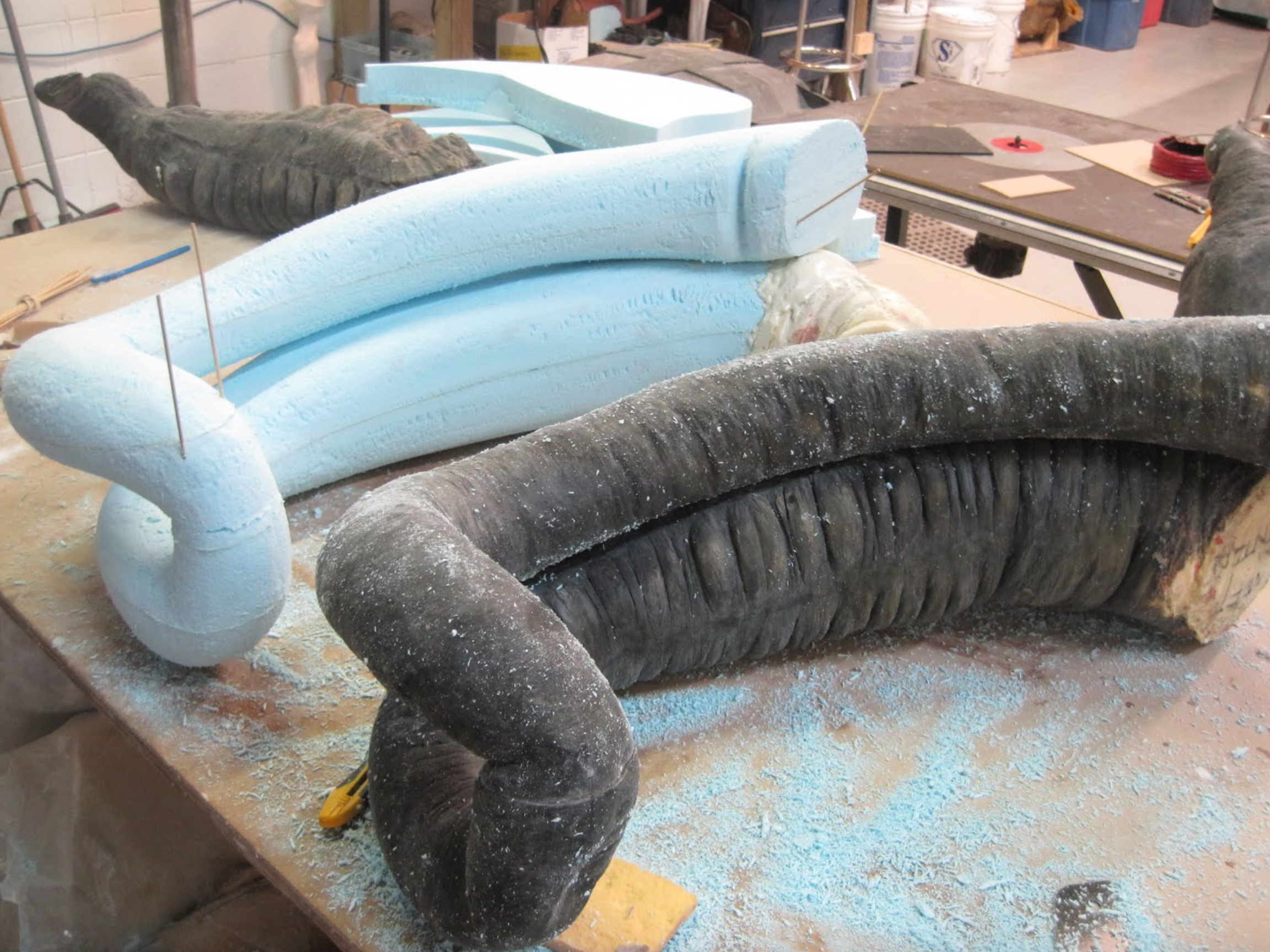
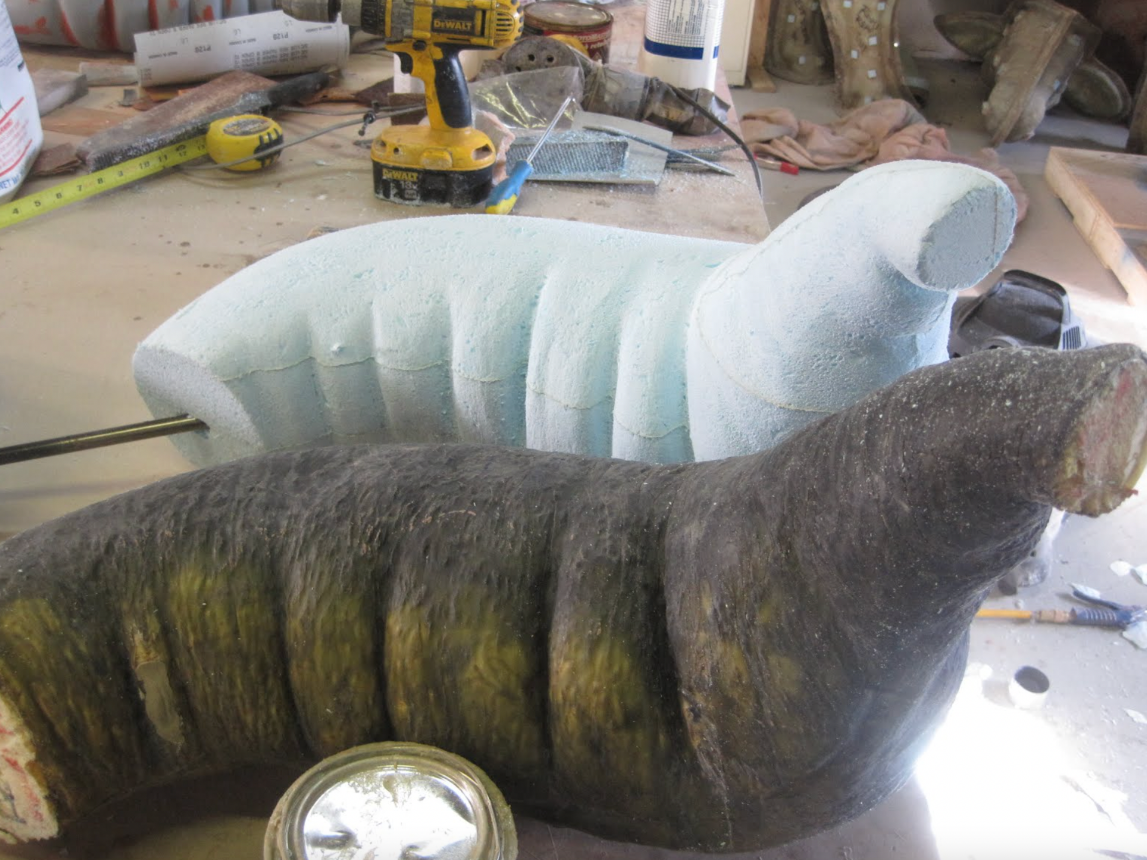
We are now beginning the refinement process for the equine colon. Reproductions of the various equine colon sections are being sculpted in styrofoam in order to improve the surface texture of the models, as well as their durability, when compared with the first prototypes. The methods of construction will also be improved (hopefully) with this re-design. This photo show the partially finished pelvic flexure, left ventral and dorsal colon.
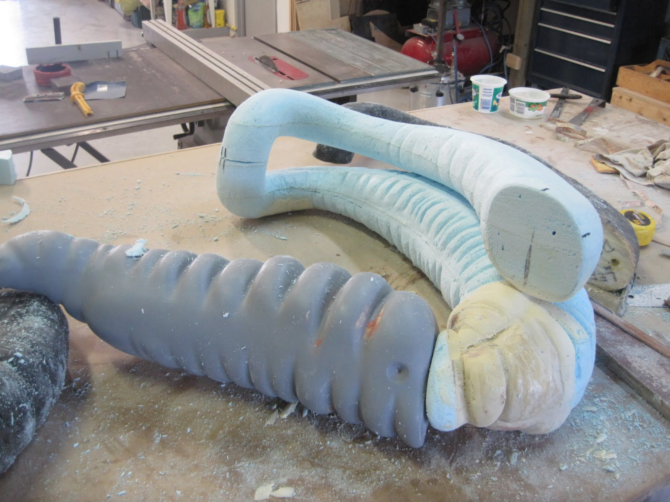
Here we are fitting the various components together as they are being sculpted. They will get an extremely smooth surface applied to them that will give a smooth realistic feel for the inflatable colon pieces. Sculptures of the transverse colon and small colon have yet to be created.
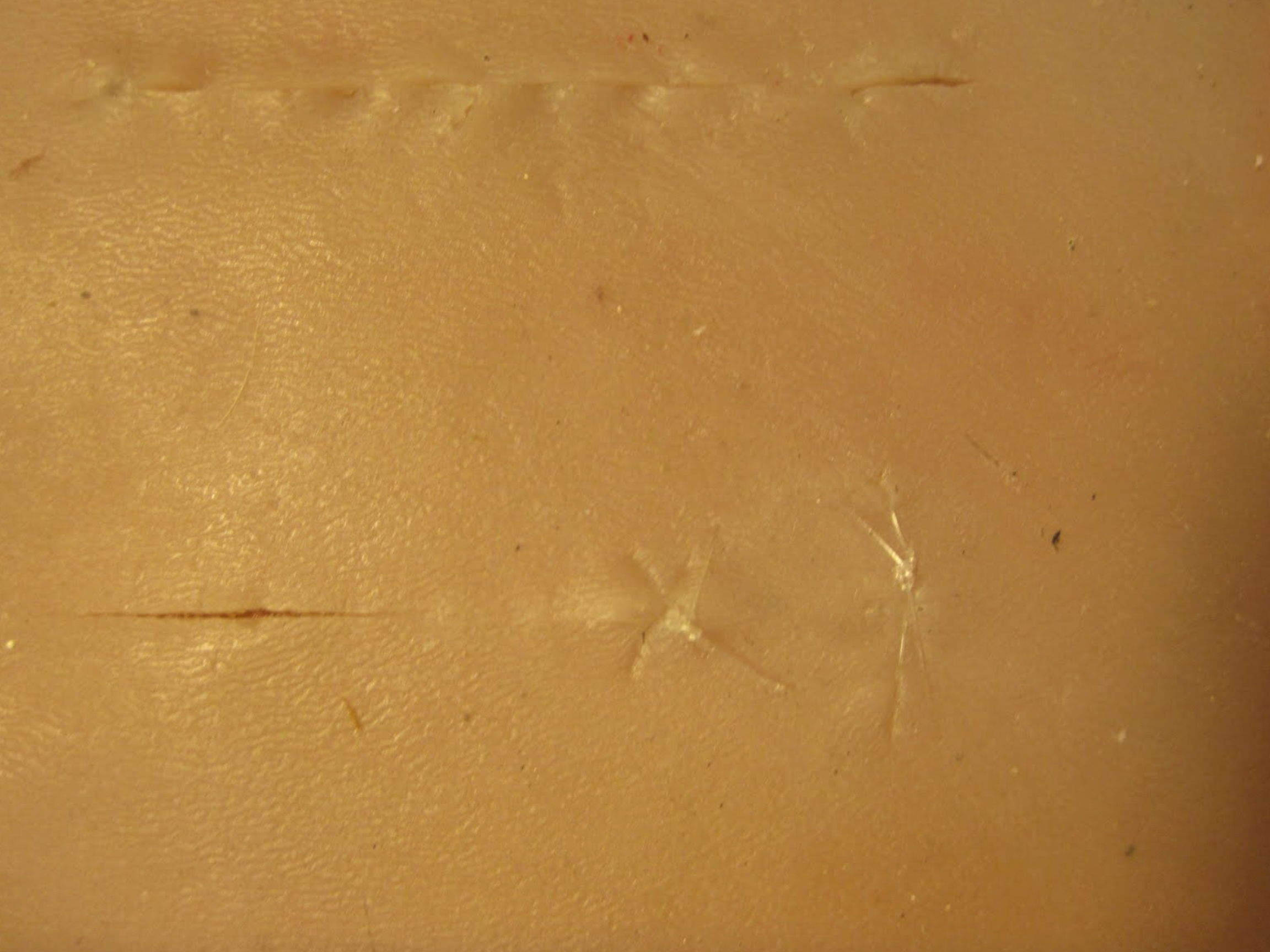
Not a very good picture of our suture pad(latest prototype). It performed very well when tested by Dr. Emma Read at then University of Calgary Faculty of Veterinary medicine. They will be tested further during OSCE’s (Objective Structured Clinical Examinations ) this week. Along with the pads which are approximately 15 cm x 15 cm , we are developing a curved platform to which the pads attach. The curvature causes the incision to open in a realistic manner. Once we confirm their functionality we can begin to produce them for anyone who may be interested.
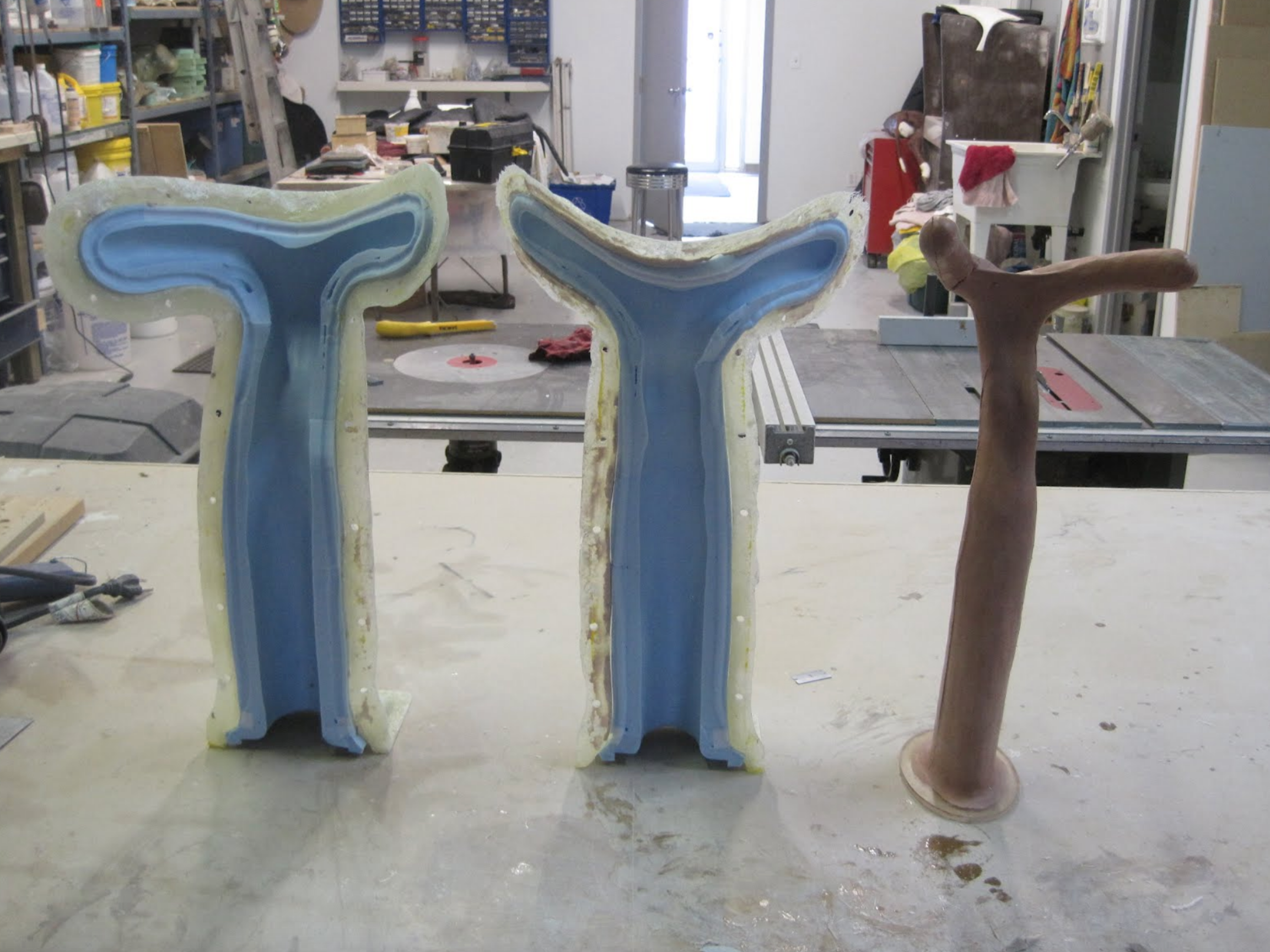
We have started development of an equine uterus model to be used in our equine model. It will be used in palpation training at UCVM. Currently biological materials are being used in the simulators, but they are difficult and time consuming to procure. When completed the uterus will have ovaries, oviducts and a representation of the suspensory ligament to psoition the uterus corrctly inside of the equine model.
A bovine uterus will also be developed, that will have several different stages of pregnancy and will be used with our bovine dystocia models.
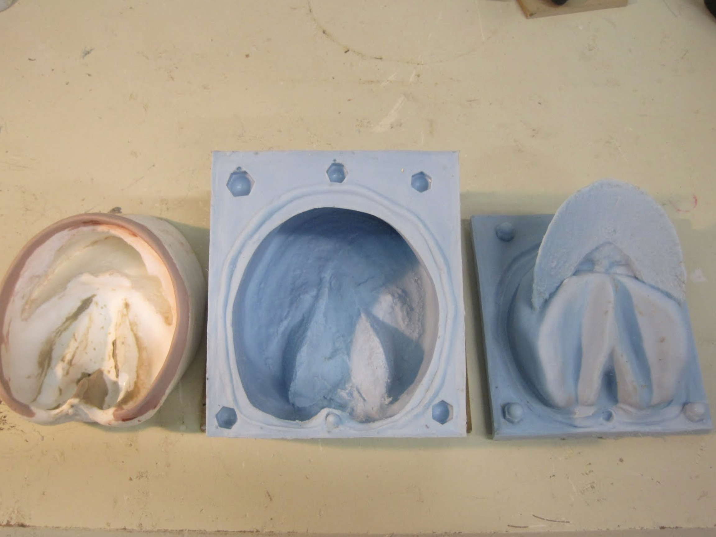
This photo shows the completed molds for the horn portion of the equine hoof. Now we will be beginning the time consuming process of sculpting the ligaments, tendons and synovial pouches of the leg. Positioning and feel of these structures is critical for palpation and injection training exercises.

This photo shows the early stages of the ligament and tendon structures of the equine leg. As we progress with the sculpting process, accuracy is ensured with frequent consultations from the UCVM faculty.

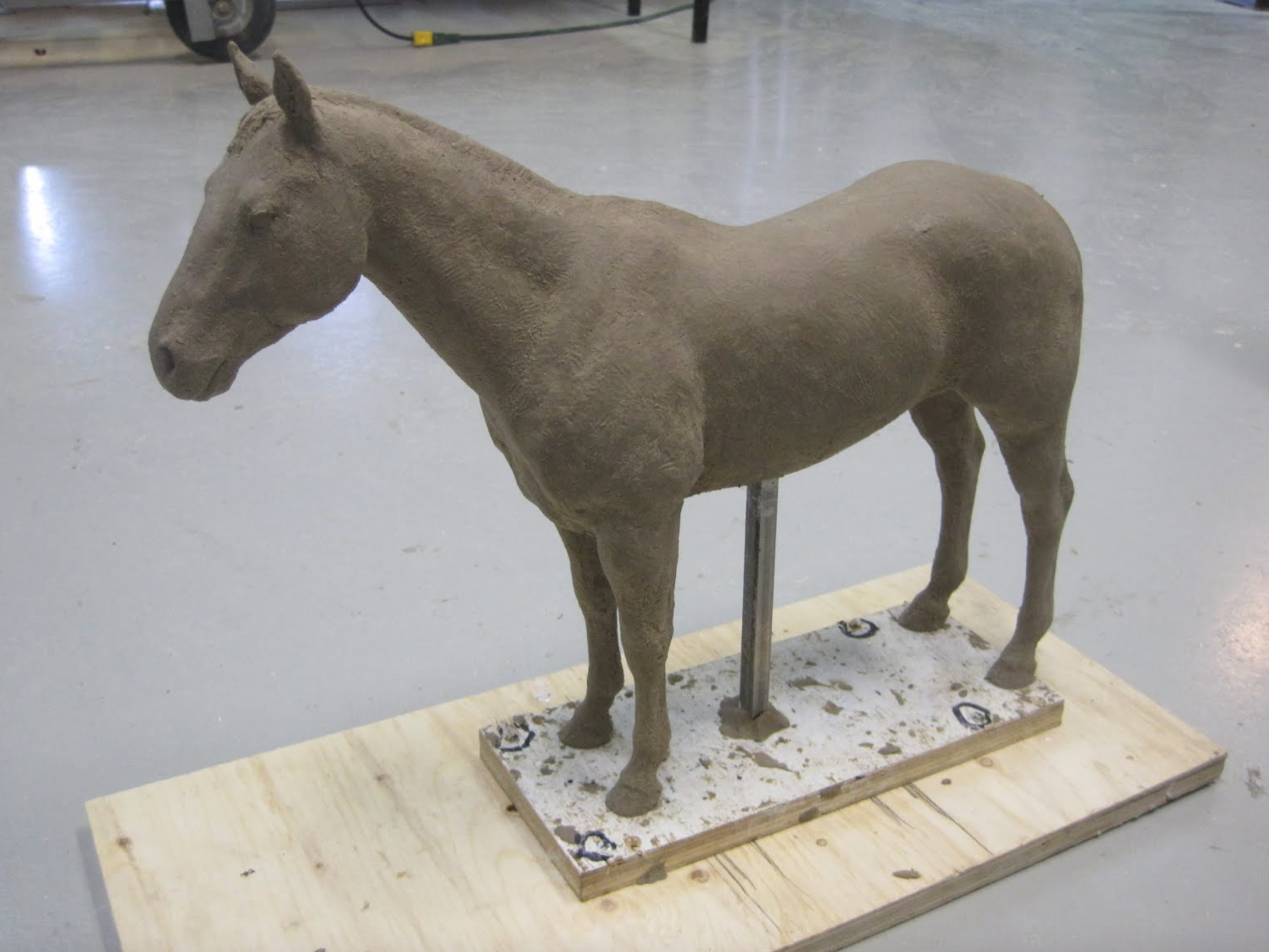
This photo shows the 1/5 scale sculpture of our new equine model. It will be the base for our colic/palpation/belly tap simulator as well as equine venipuncture simulator. It will be reproduced in full scale, then molded and reproduced. The model was sculpted by Al Stinson from a live model.
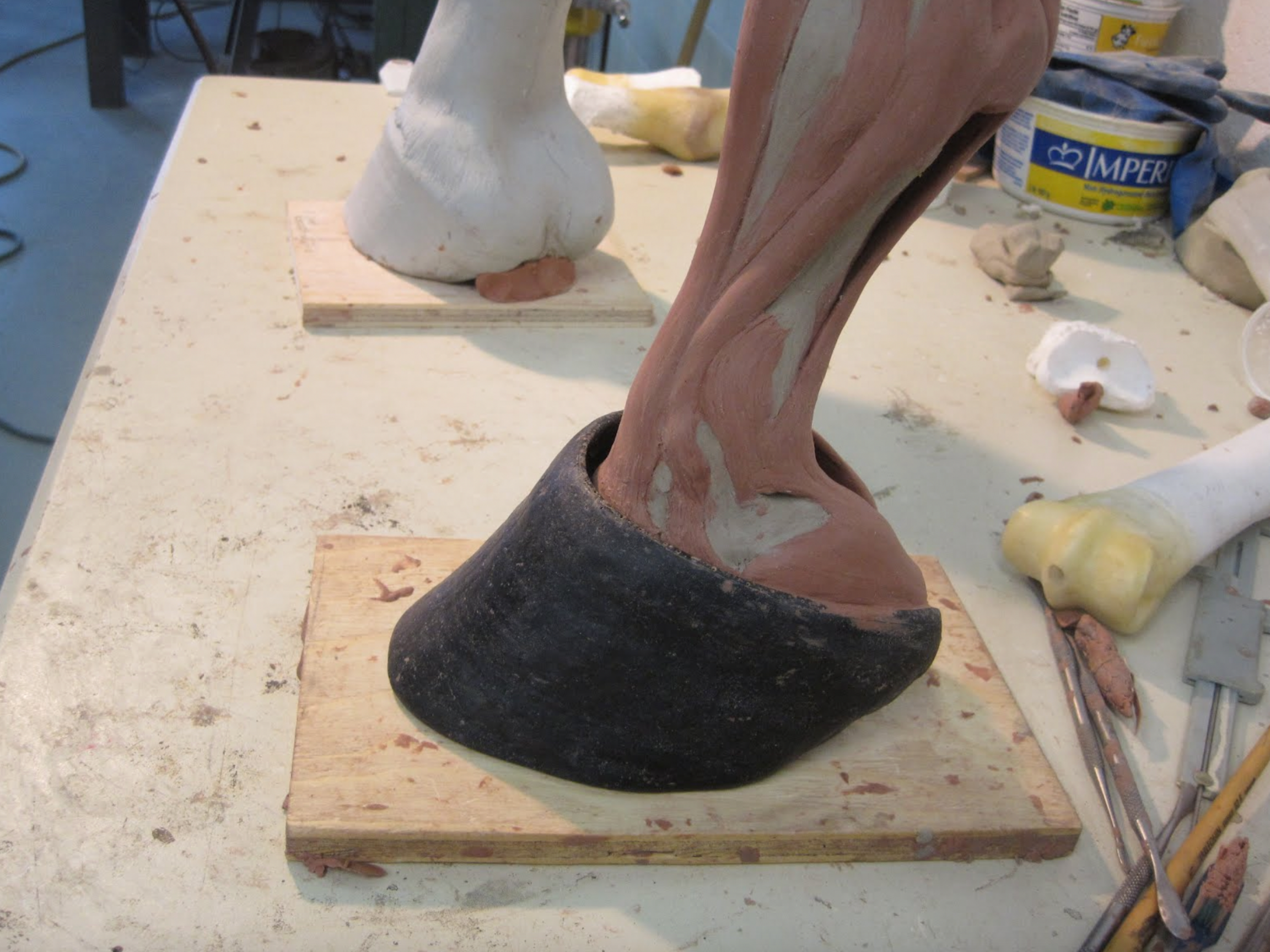
The sculpture of the tendons and ligaments is near completion. throughout the entire sculpting process we have been consulting with Dr. Emma Read and using many anatomical text books in order to ensure a high degree of accuracy.

Dr. Emma Read describes structures of the equine foreleg.

Photo of completed tendon/ligament structure. Prototype unit is now ready to receive the skin. Several different materials wil be tested in order to get the optimal results.


The equine uterus is progressing with a few size changes being made. We have re-sculpted the model to reflect those changes. , deemed appropriate by a group of equine reproduction specialists. Here we see the cast of the new model and the mold for it. We will be creating various ovaries to represent different stages of pregnancy along with a broad ligament to support the uterus in our equine model.

The equine injection leg prototype is nearing completion. The leg contains all the appropriate structures for determining proper needle placement. Fluid can be drawn and injected from and into the synovial pouches in the joints. The procedure can be repeated multiple times. The number of times this can be accomplished without degradation of the skin will be determined with further testing.


Front view of the leg. All the joints can be flexed to facilitate injection where necessary.
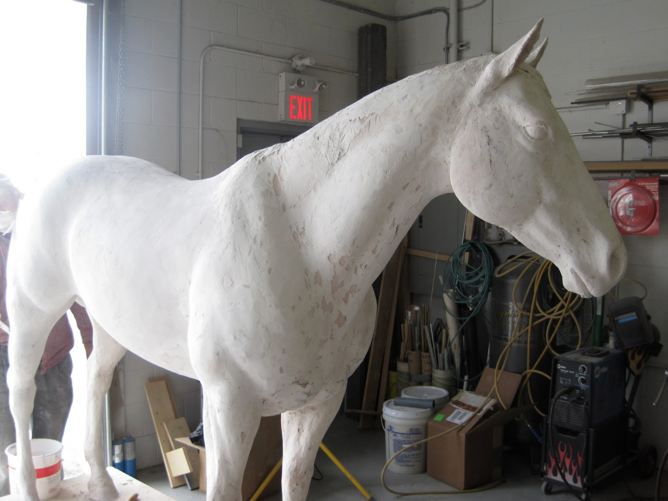
Our new Quarter Horse model takes shape. This will be the basis for our equine colic simulator as well as our equine venipuncture simulator. By creating our own model we are able to ensure the quality of the finished fiberglass inside and out, as well as the anatomical accuracy.
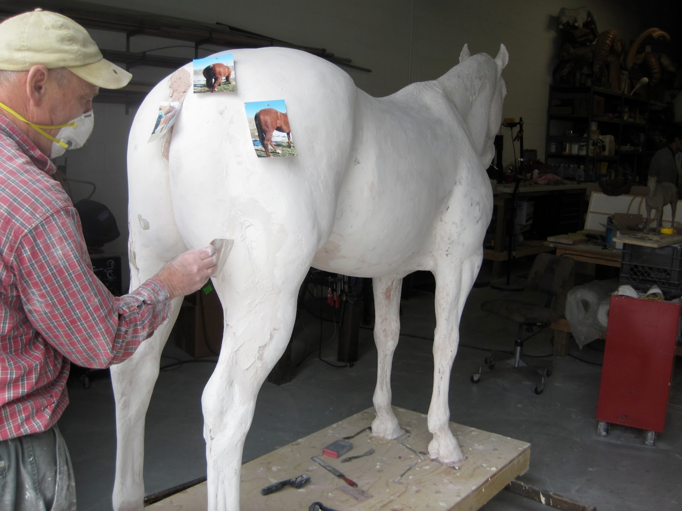
Al Stinson refines the structure with help from photos and working with live models. The base of sculpture is a foam CNC cut reproduction of a 1/5 scale model that Al created. It was laser scanned then enlarged to actual size. Refining is necessary to ensure accuracy that cannot be achieved in the small scale.

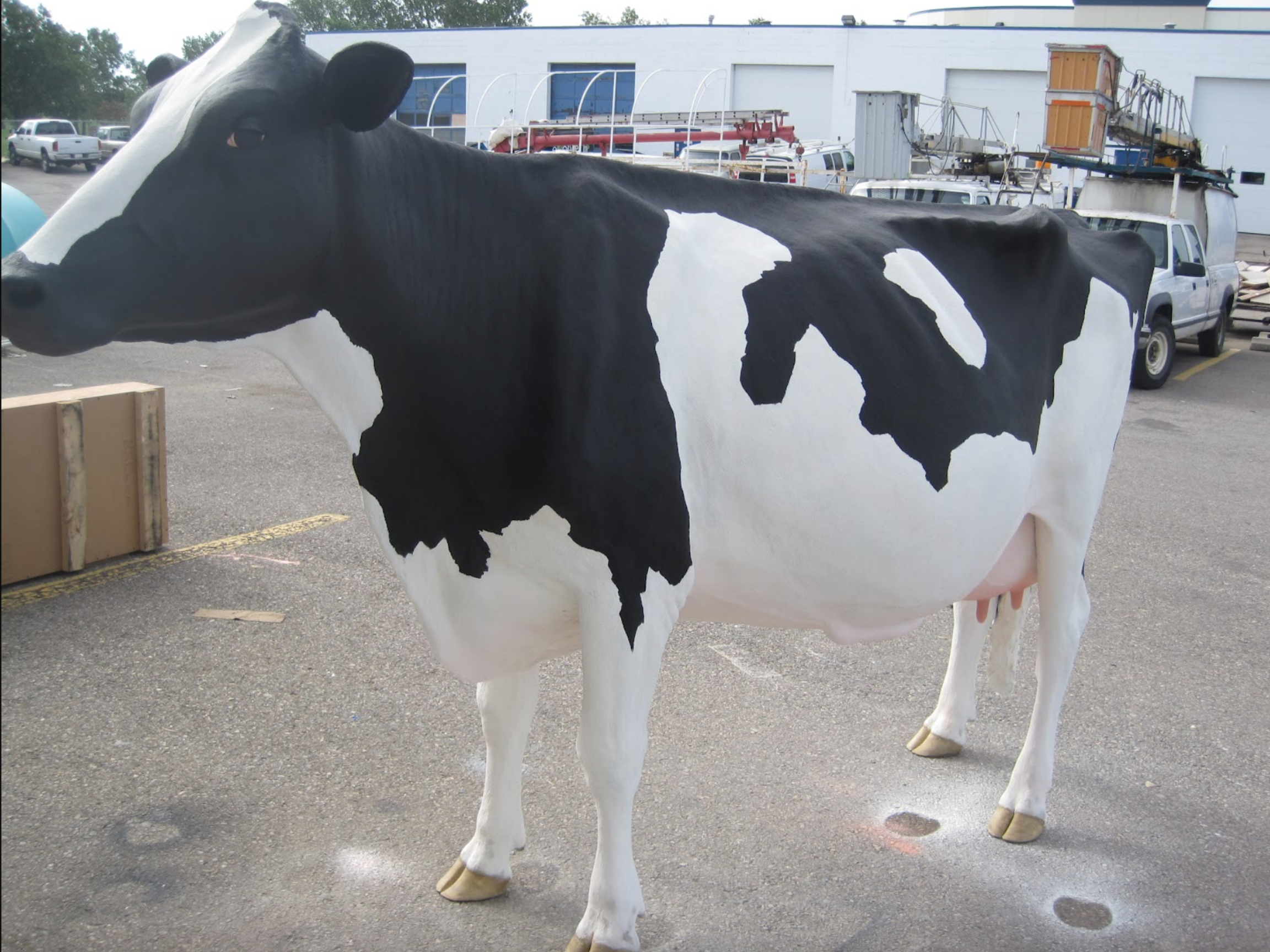
This Holstein model is completed and awaiting pick-up. Alberta Milk will be using it at their display at the Calgary Stampede to demonstrate production milking equipment.
To facilitate this the model has soft realistic teats, that are easily replaced.
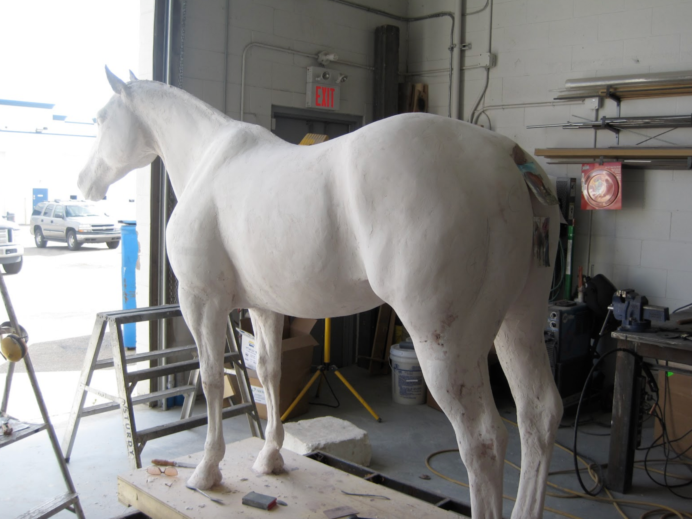
The finished Quarter Horse sculpture is ready for the molding process. This will be the basis for our equine palpation simulator as well as the equine venipuncture model. At some point we may combine them in one model.
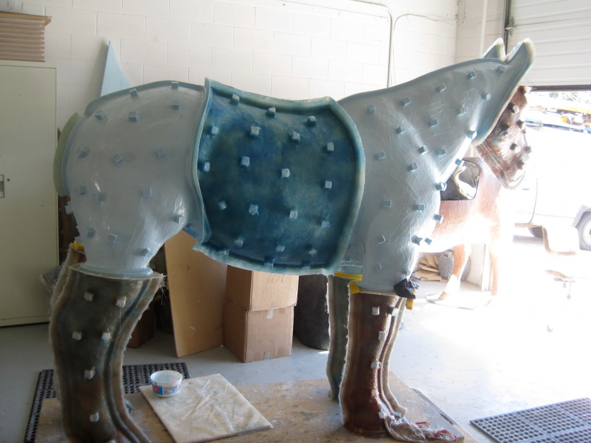
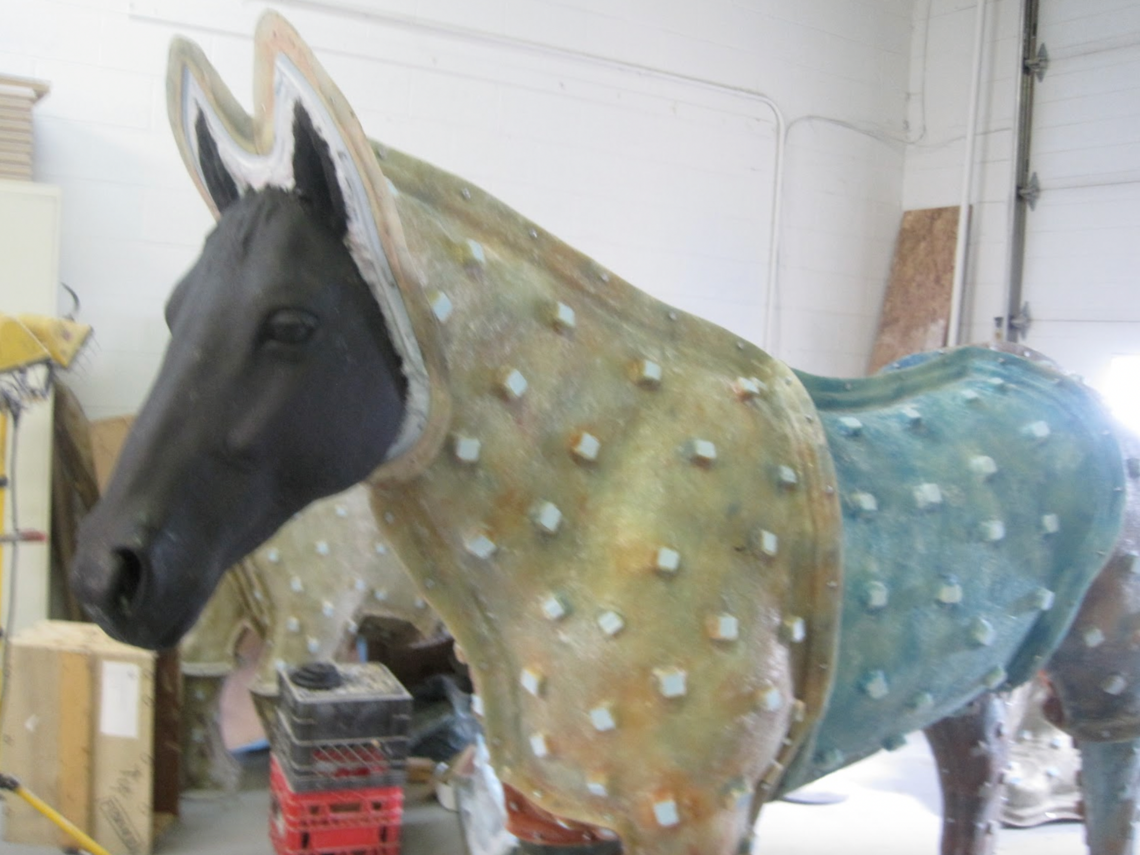
The Quarter Horse mold has been completed and the first fiberglass model begins to emerge from it. Once complete all the internals will be added (cecum, right and left ventral colon, right and left dorsal colon,section of small intestine, left kidney, spleen, uterus, ovaries) along with soft perineum panel, tail, pelvis and belly tap function.
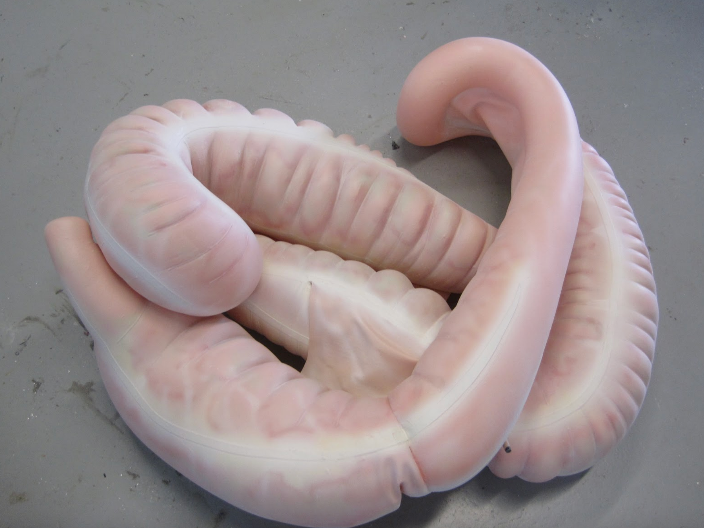
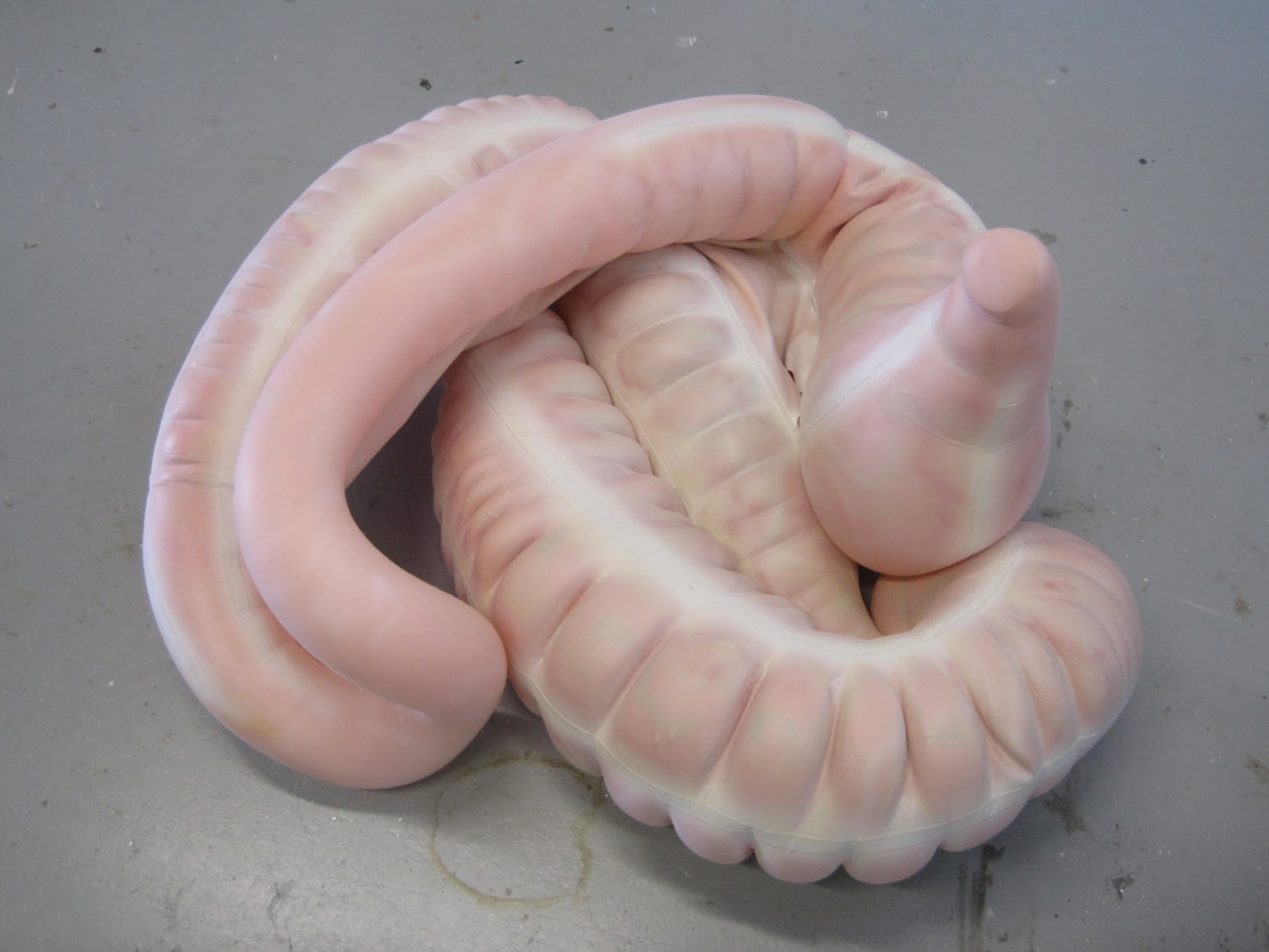
The next two photos show the completed inflatable colon model that will be used in the equine palpation/colic model. The colon can be positioned to simulate various colic conditions and displacements.
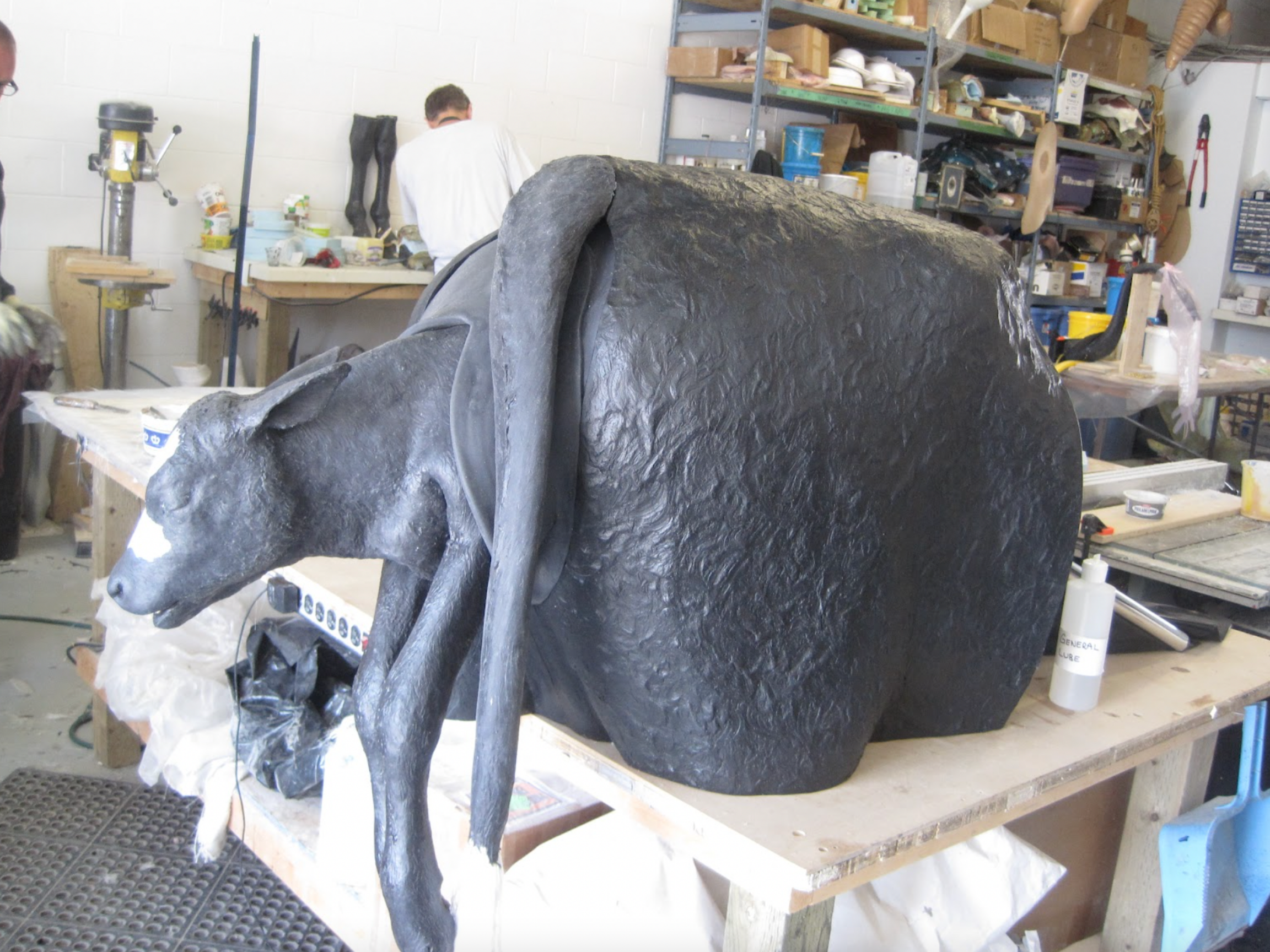
This is a prototype table top bovine dystocia unit being developed for Olds Agricultural College, in Olds, Alberta. It consists of the rear quarter of the body with pelvis, uterine bag, pneumatic support, soft perineum panel and tail. It will be tested and evaluated at Olds College and then we will make any changes and improvements that are necessary.

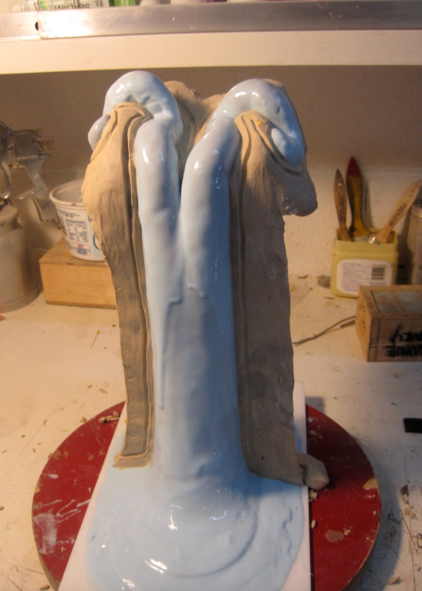
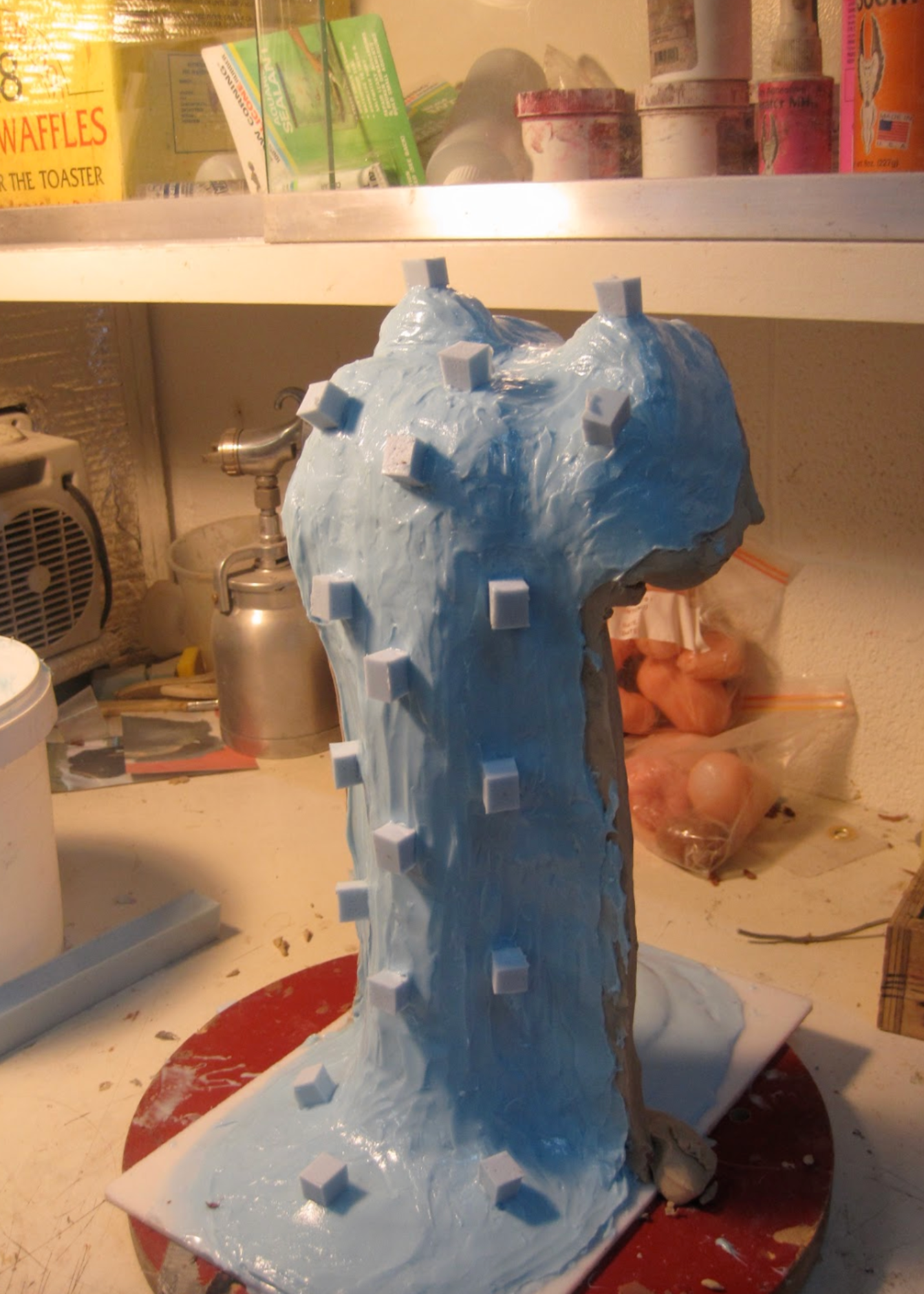
The rubber detail layer has been completed at this stage and the fiberglass mother mold has been started.

This photo shows our new quarter horse model. This particular unit is heading to Colorado State University.


Another view of the equine colic simulator. This unit has an inflatable GI tract(right and left ventral colon, right and left dorsal colon, cecum , and portion of small intestine). It also has representations of left kidney, reno-splenic ligament and spleen, along with a pelvis and inflatable section of rectum, soft perineum panel and tail. A belly tap function is also included.
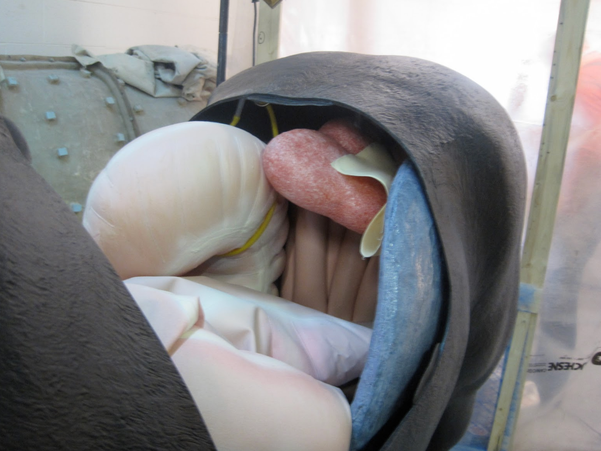
Here we can see the GI tract, small intestine, spleen and kidney
This photo shows the inflatable portion of the rectum and our newly developed equine uterus with broad ligament. The uterus also has interchangeable ovaries for palpation training
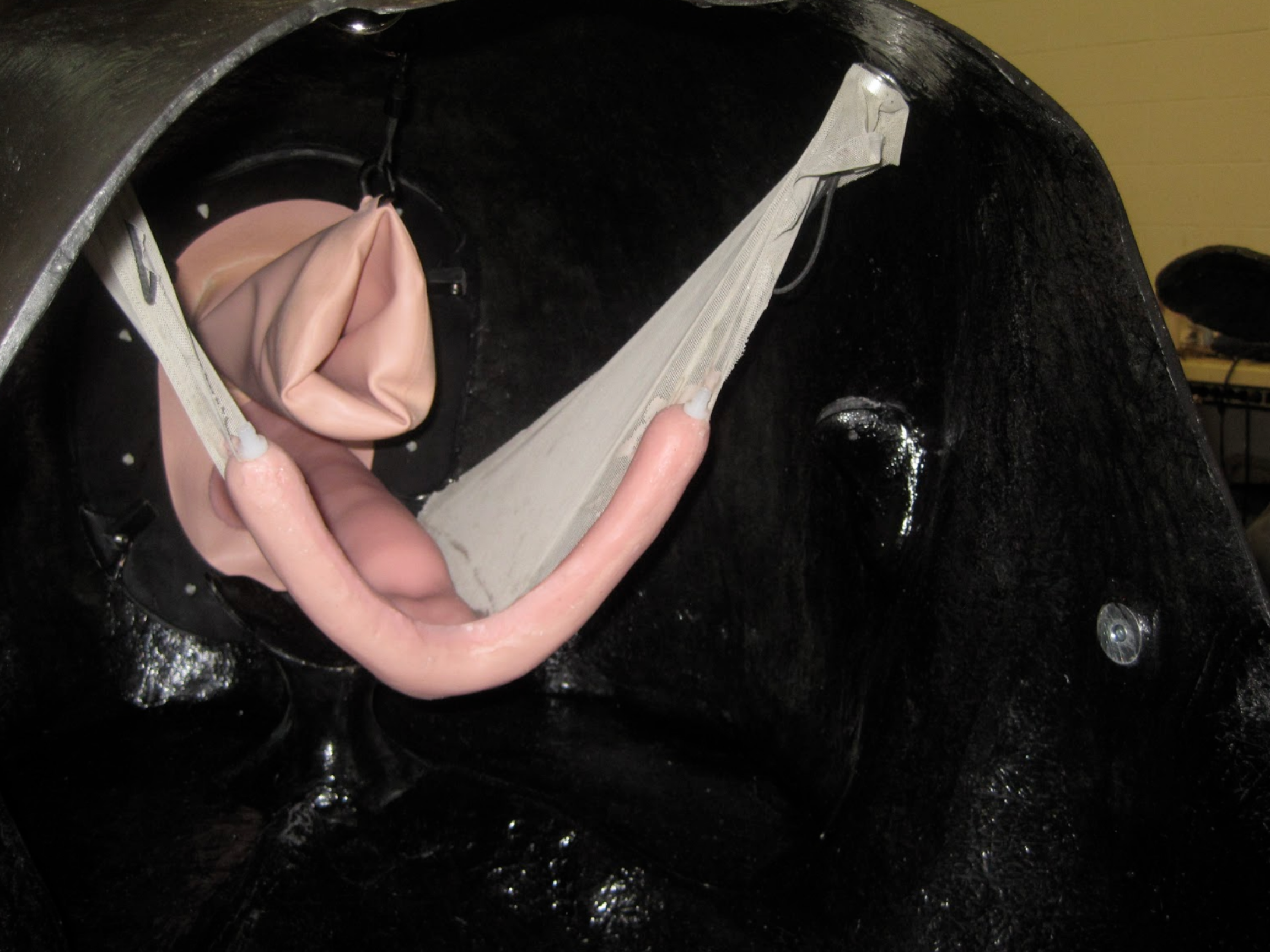
Rear view of the new quarter horse model.

Once completed the model will be molded and reproduced in hand laid epoxy/fiberglass.
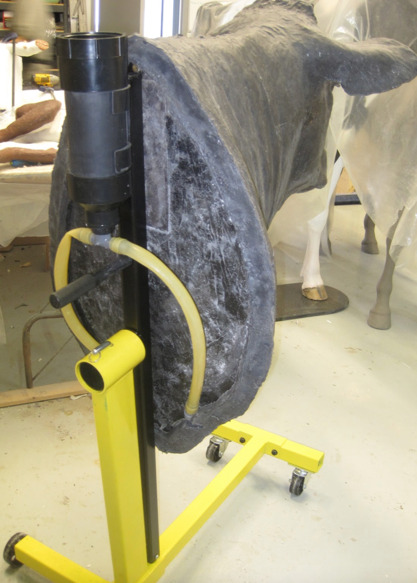
The jugular vein is connected to a tank that creates a small amount of pressure in the vein. The vein itself can be replaced once it has had multiple punctures.
Once we have had the unit tested and have made any changes necessary we will create a dairy cow version and an equine version.
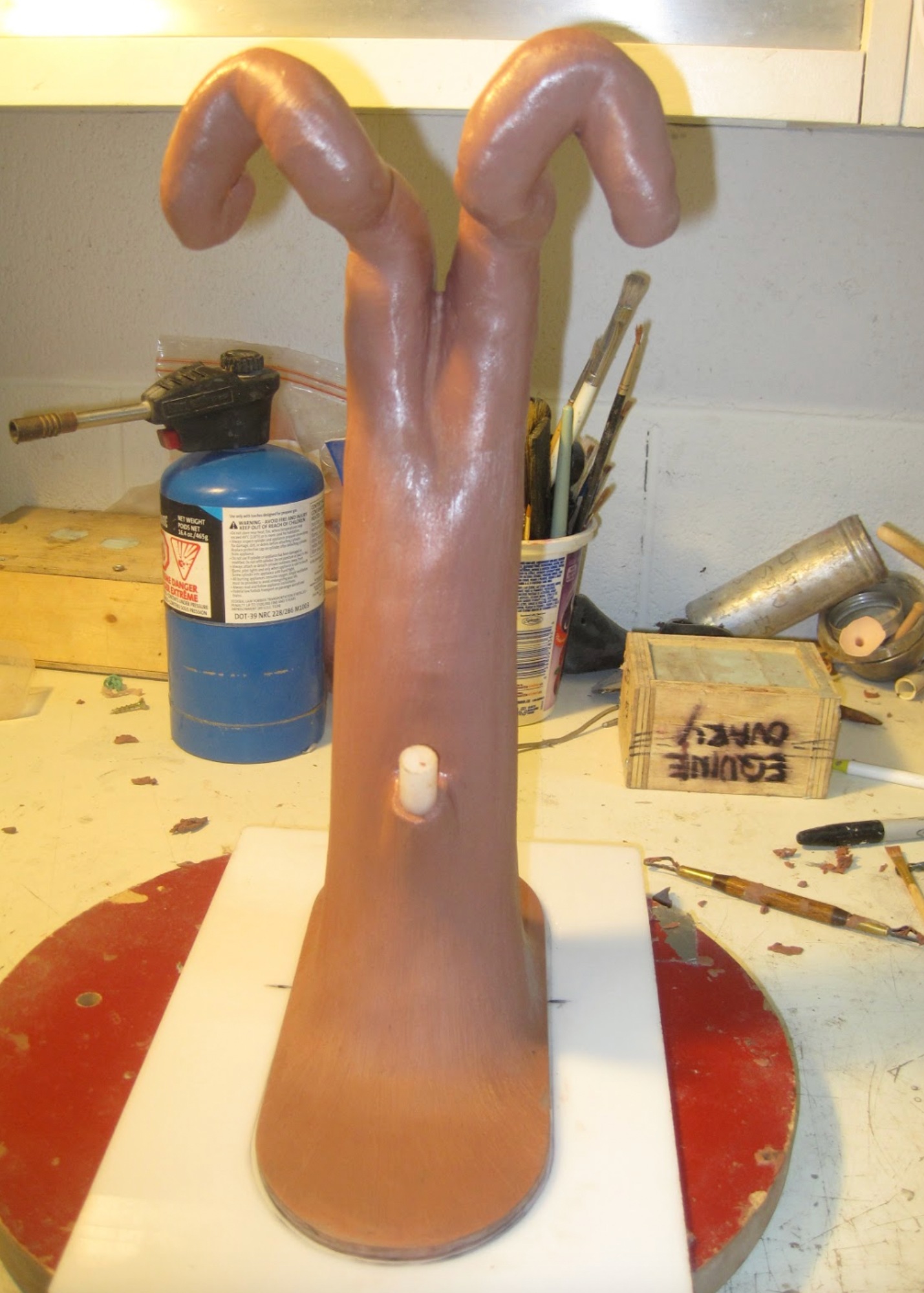
This photo shows the original sculpture of the bovine open uterus model. We will create additional sculptures for later stages of pregnancy as well as creating a uterine slip for early pregnancy detection training purposes.
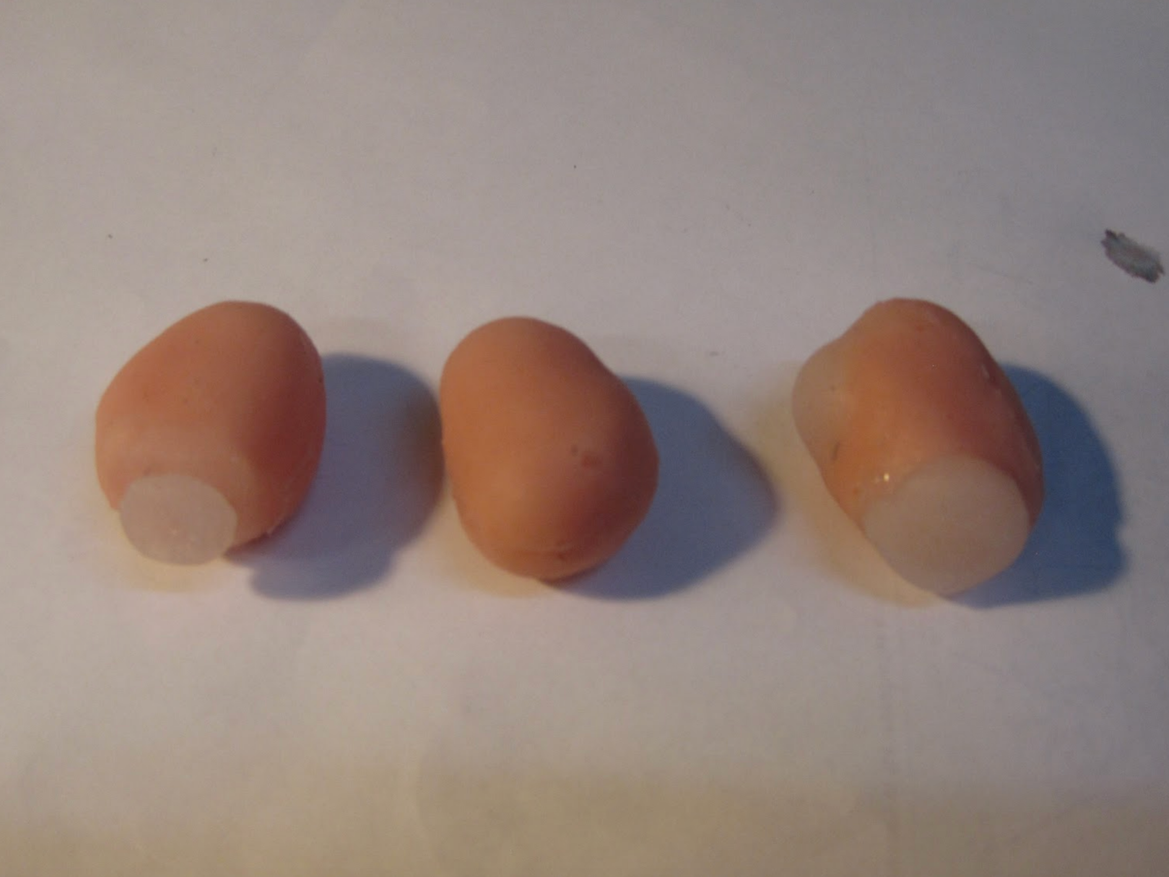
This photo shows the various interchangeable ovaries, with CL and follicles.
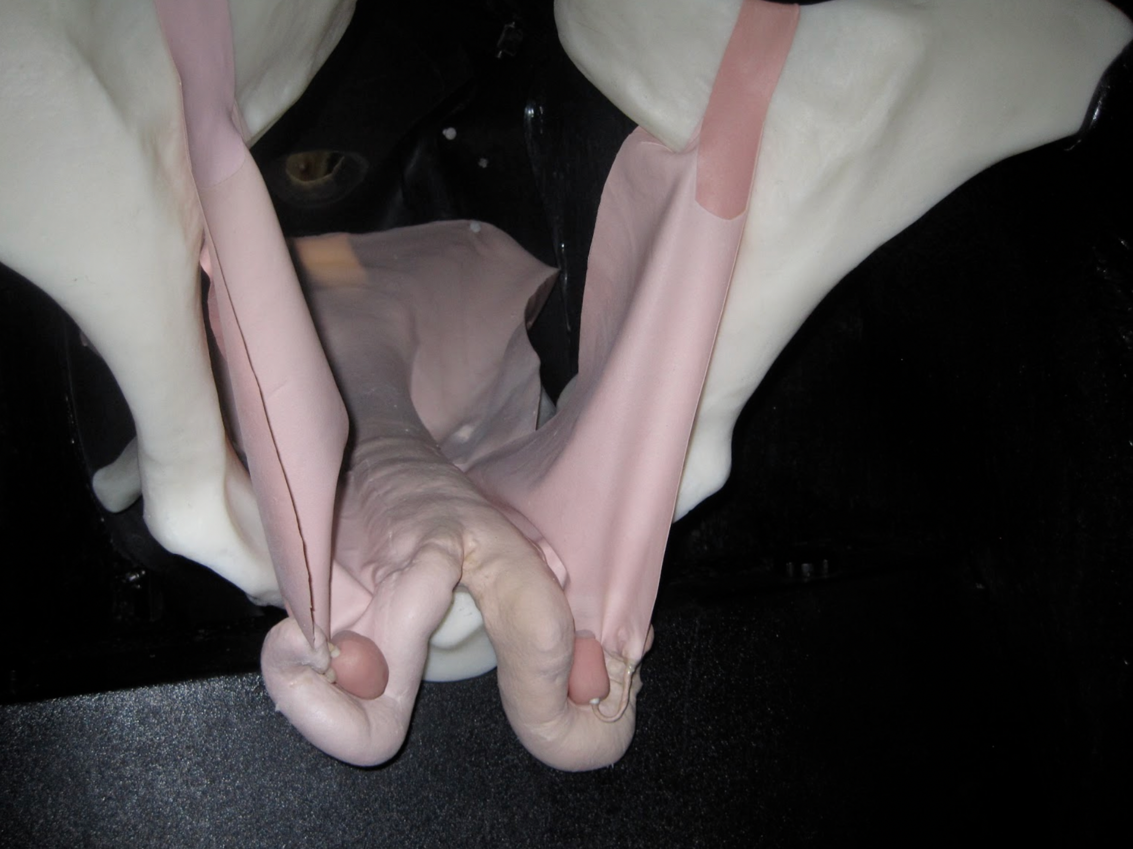
Here we have the bovine open uterus suspended in the pelvis along with the broad ligament and ovaries. The uterus is easily exchanged for uterii with varying stages of pregnancy.
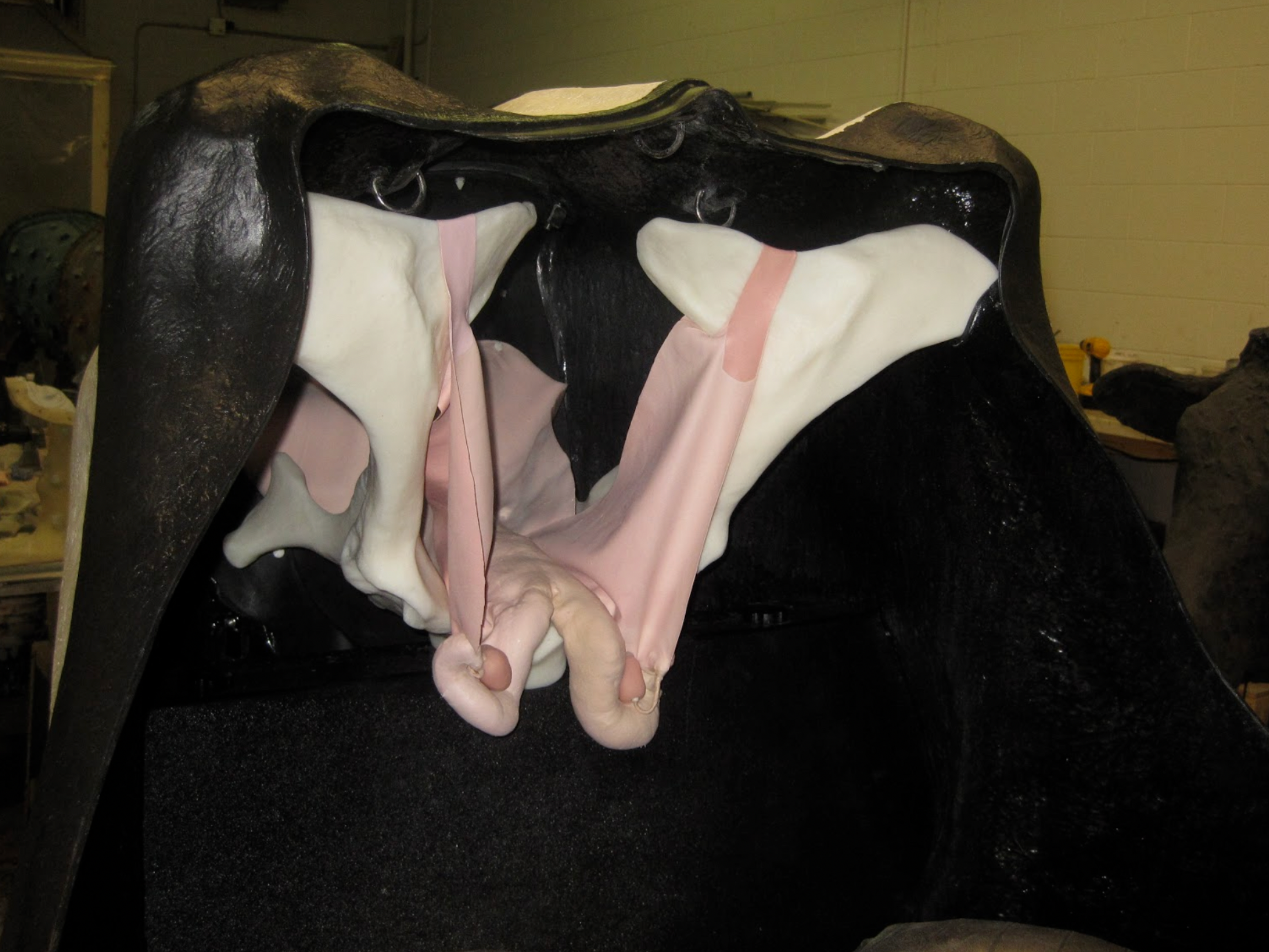

These next 4 photos show a prototype equine tendon/ligament model. It was created as part of the development process for the equine injection model. Also as part of that process a forelimb skeletal model was developed.
Both the skeletal model and ligament/tendon model are cast in a rigid plastic.
We will be continuing development of the injection limb along with a variety of companion teaching models.


UCVM student performing a uterine swab on the equine simulator. Actual equine reproductive tracts were hung in the simulators for the OSCE’s. We are currently testing a simulated equine uterus to replace the use of biological materials for this procedure.
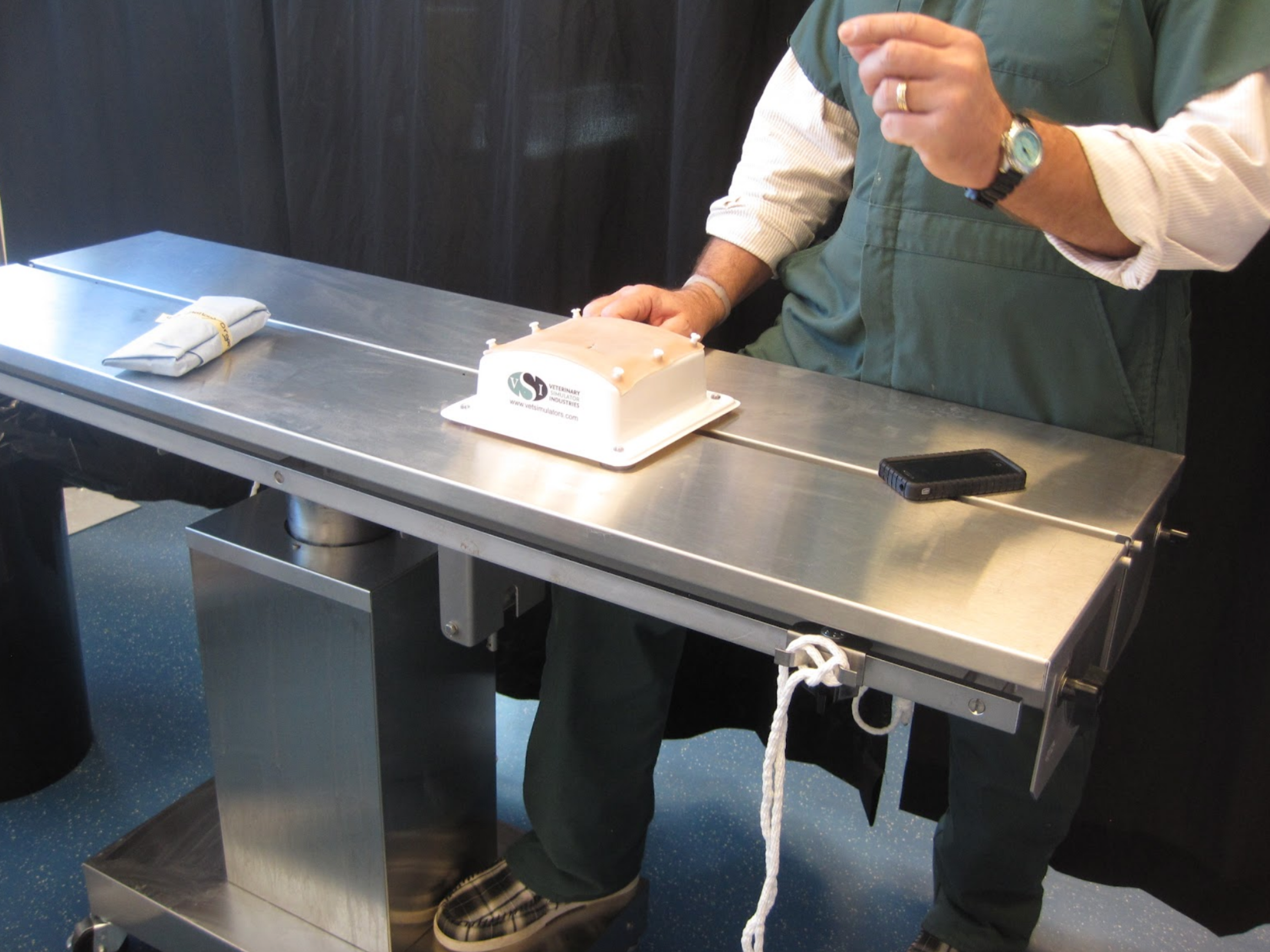
Also used at UCVM during the OSCE’s were our hollow organ suture training pads. They functioned well and provided a very realistic feel and function.
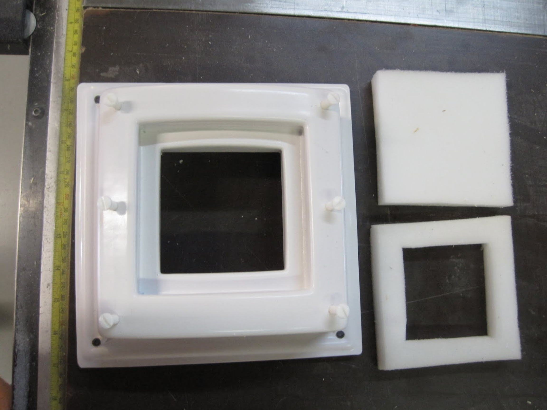
This the prototype base unit for the suture/hollow organ pads. Both pads can be used with the same base by exchanging the foam inserts. The unit has suction cups on the bottom to prevent movement while suturing.
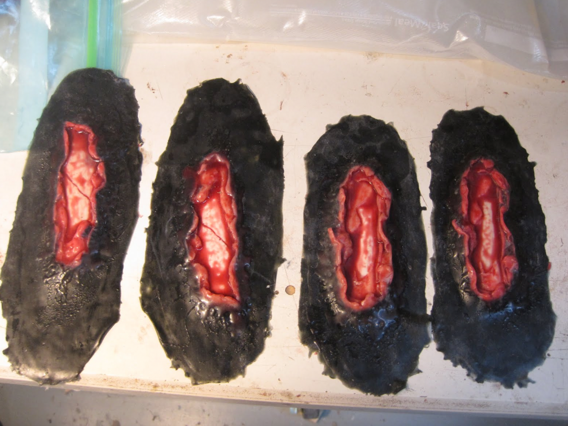
We have created simulated equine leg wounds for accident triage training. these wounds can be applied to the leg and re-used multiple times. UCVM does simulated horse trailer accidents for students and veterinarians.
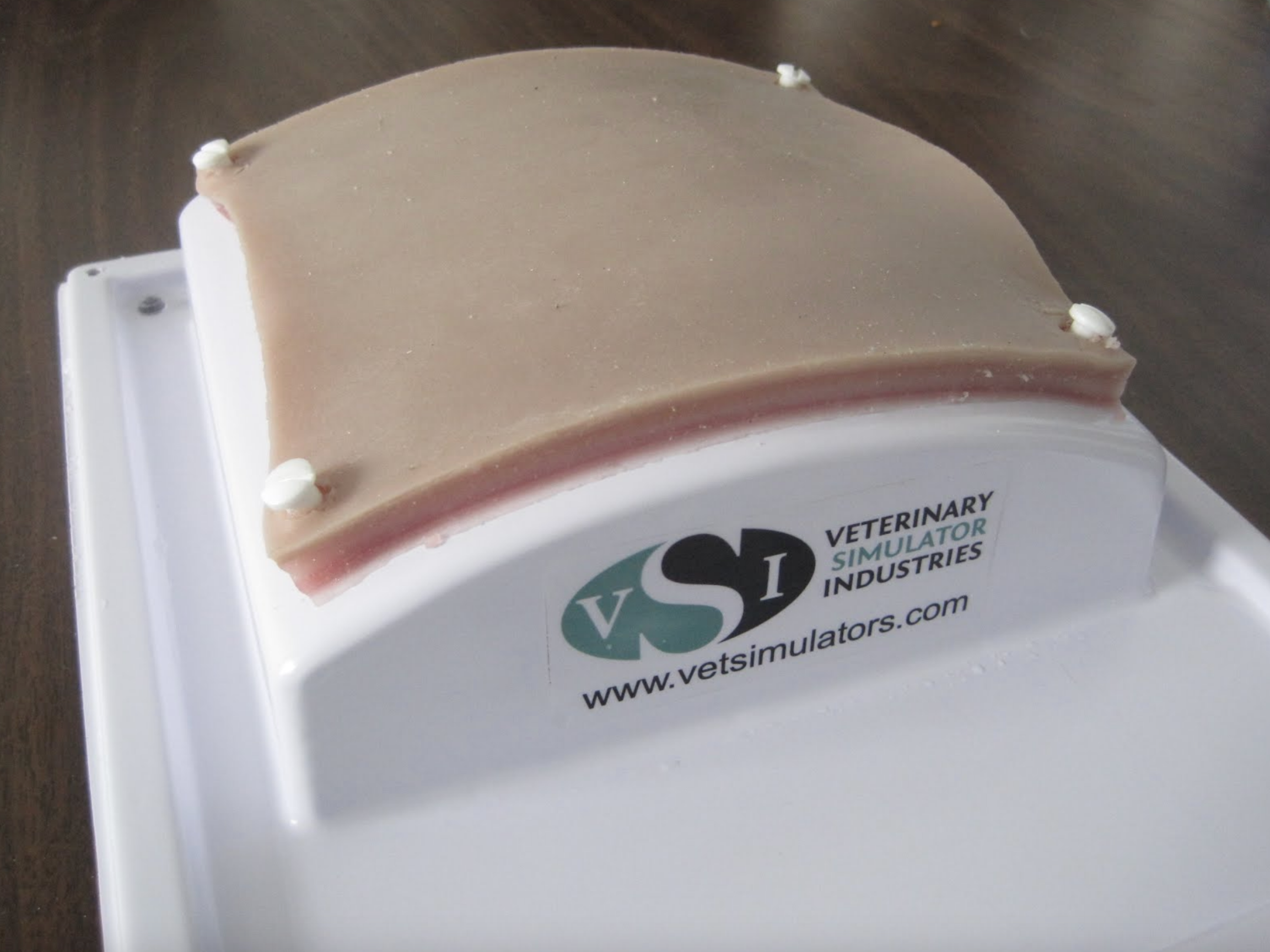
One of the recent suture pad prototypes, with its base. the base has suction cups for stability and can accept both the 4 layer suture training pads and the hollow organ pad.
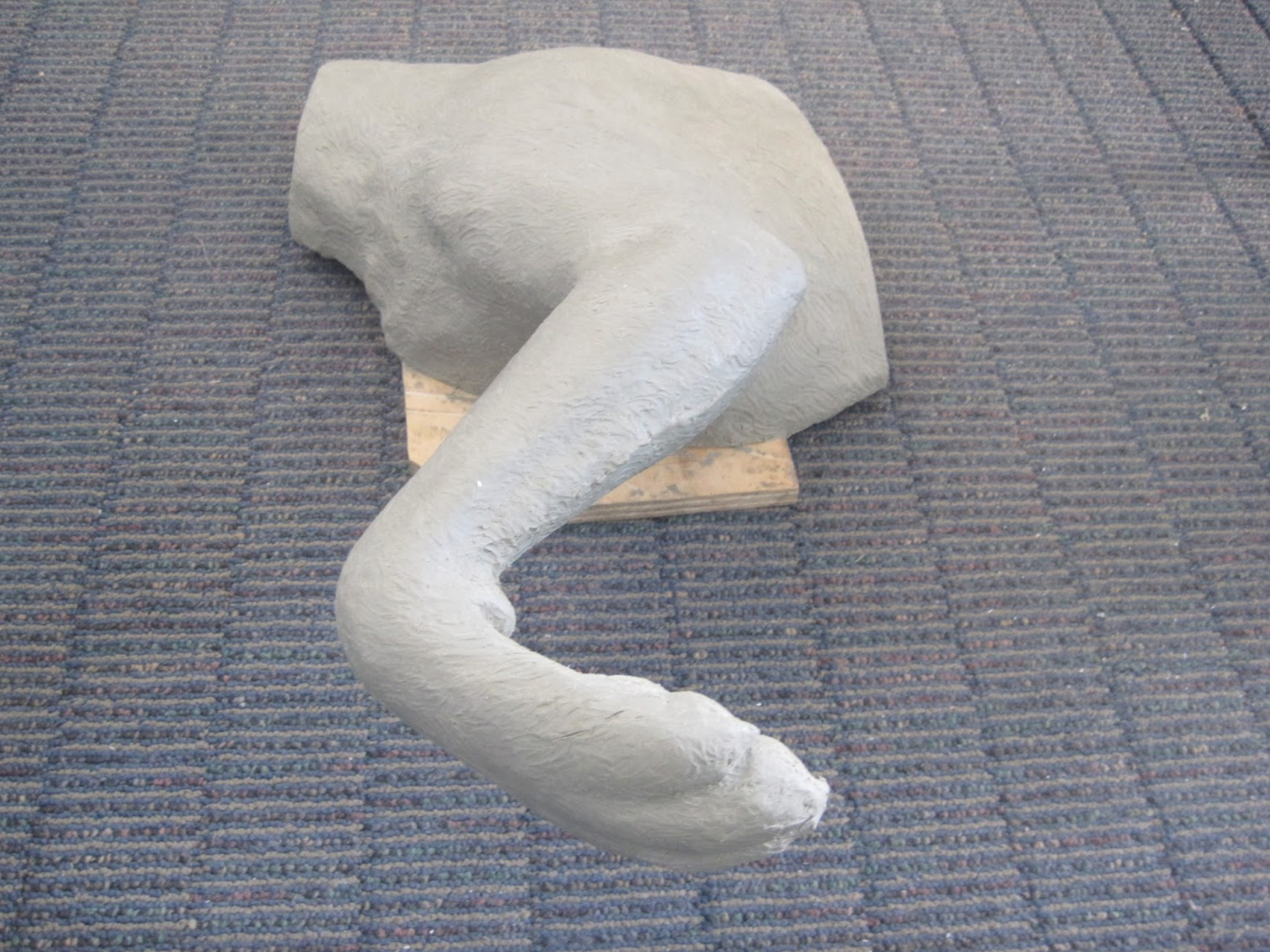
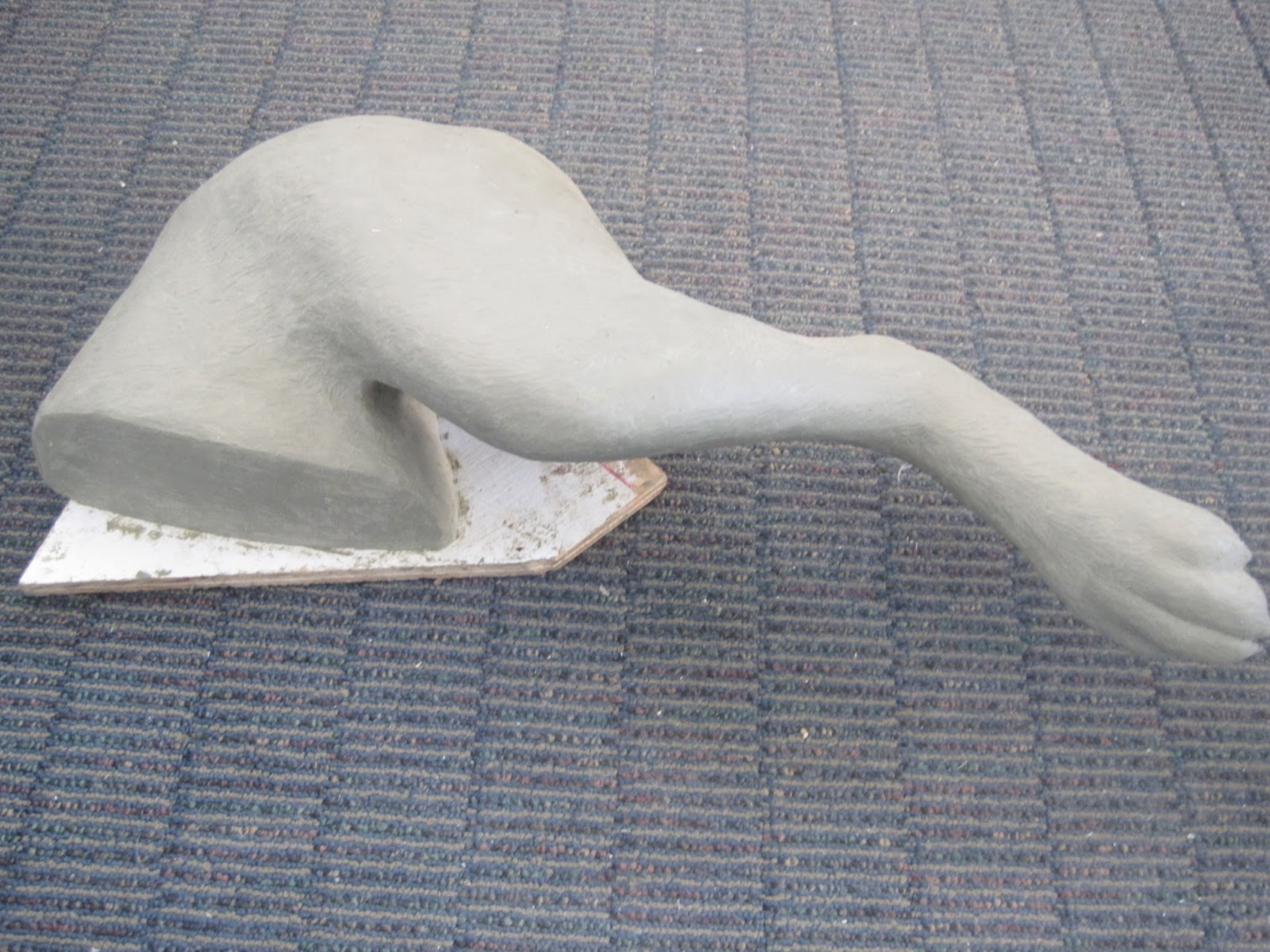
Original clay sculptures of both front and rear canine limbs. (Labrador). These were used to create articulated limbs to be used in bandaging/sling training at Ross University.
The fully articulated limbs were created with steel skeletons and rubber skins, along with mounting points to facilitate attachment to tables.

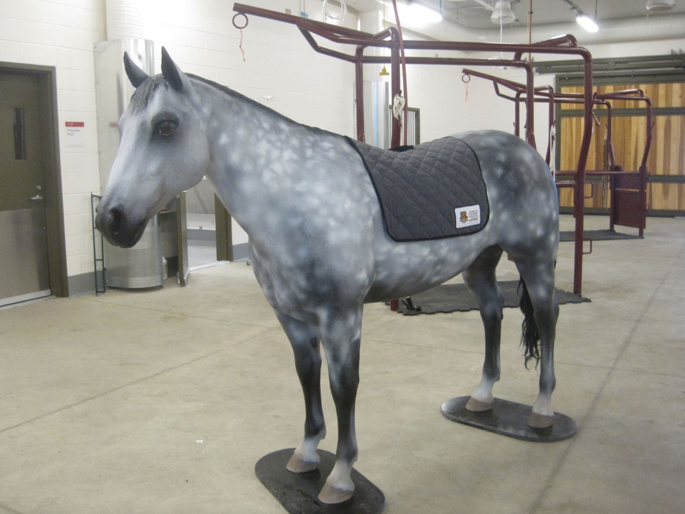
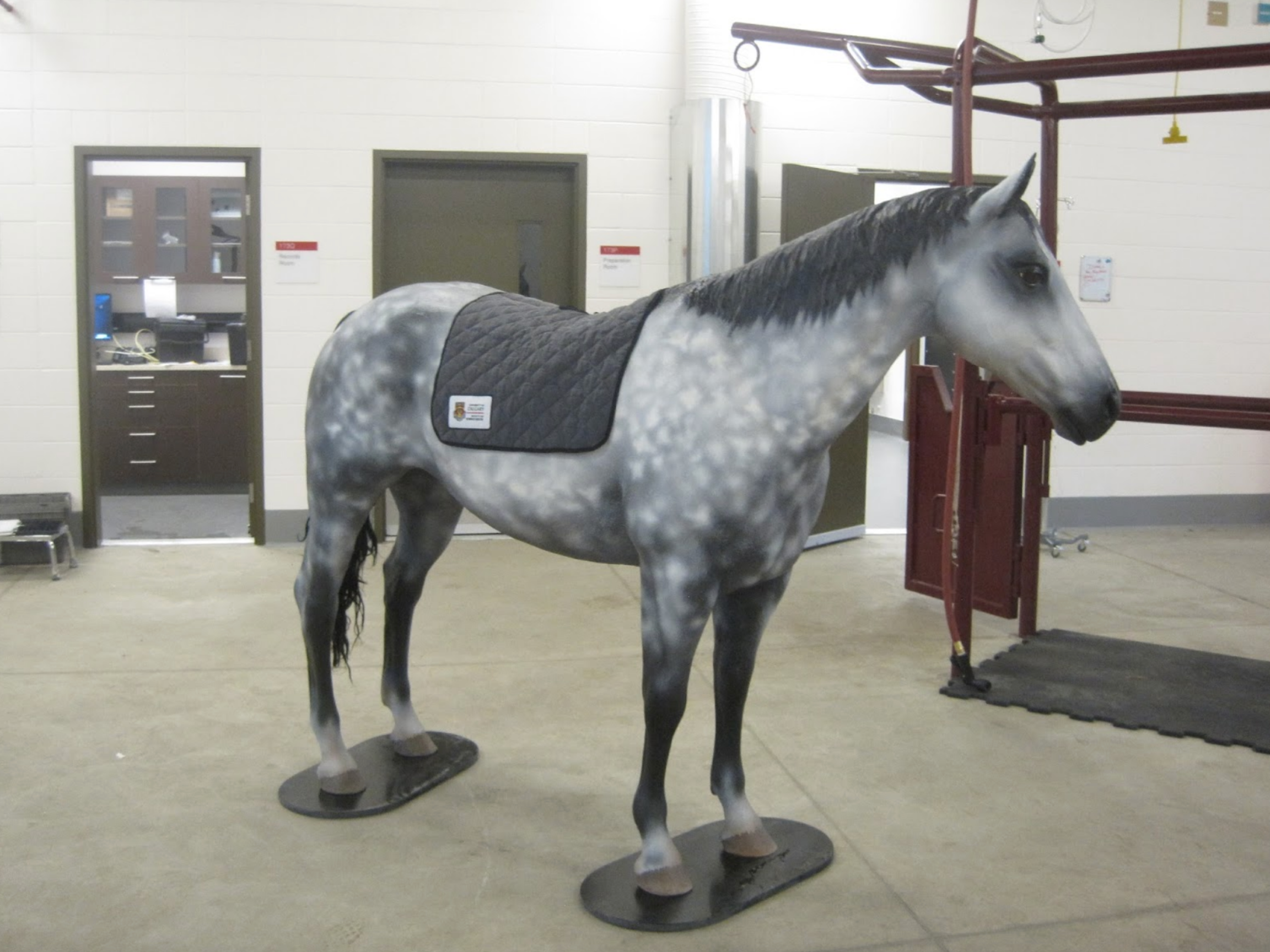
This is our new Quarter Horse model specially painted as a Dapple Gray breed for Dr. Emma Read of the University of Calgary Faculty of Veterinary Medicine.
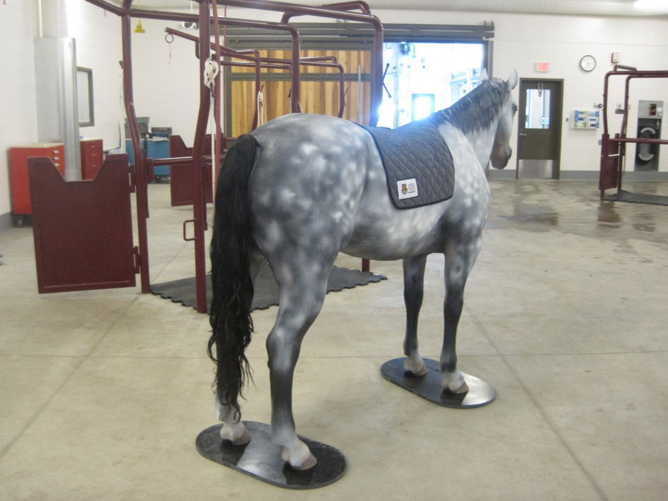
This particular simulator has the standard features of this model, inflatable GI tract for colic simulation, abdominocentesis function, pelvis and soft perineum panel, but also has a prototype reproductive tract that can be used for uterine biopsy simulations and training.
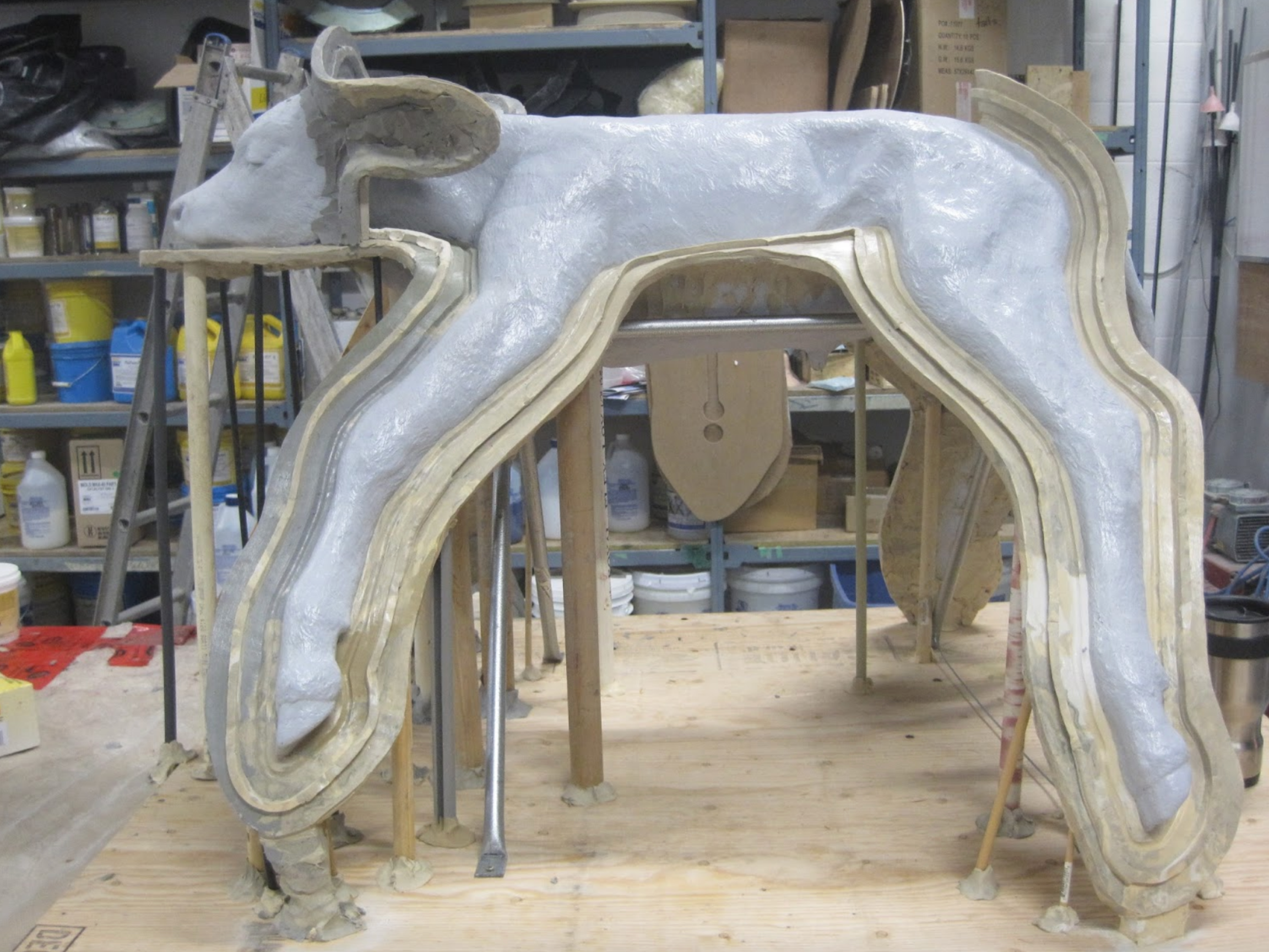
Here the dystocia calf model is being prepared to create a new mold. Improvements are being made during this process that will improve the quality of the finished product.

The dystocia calf model part way through the molding process. We have made improvements to both sculpture and mold to improve quality of the finished product.
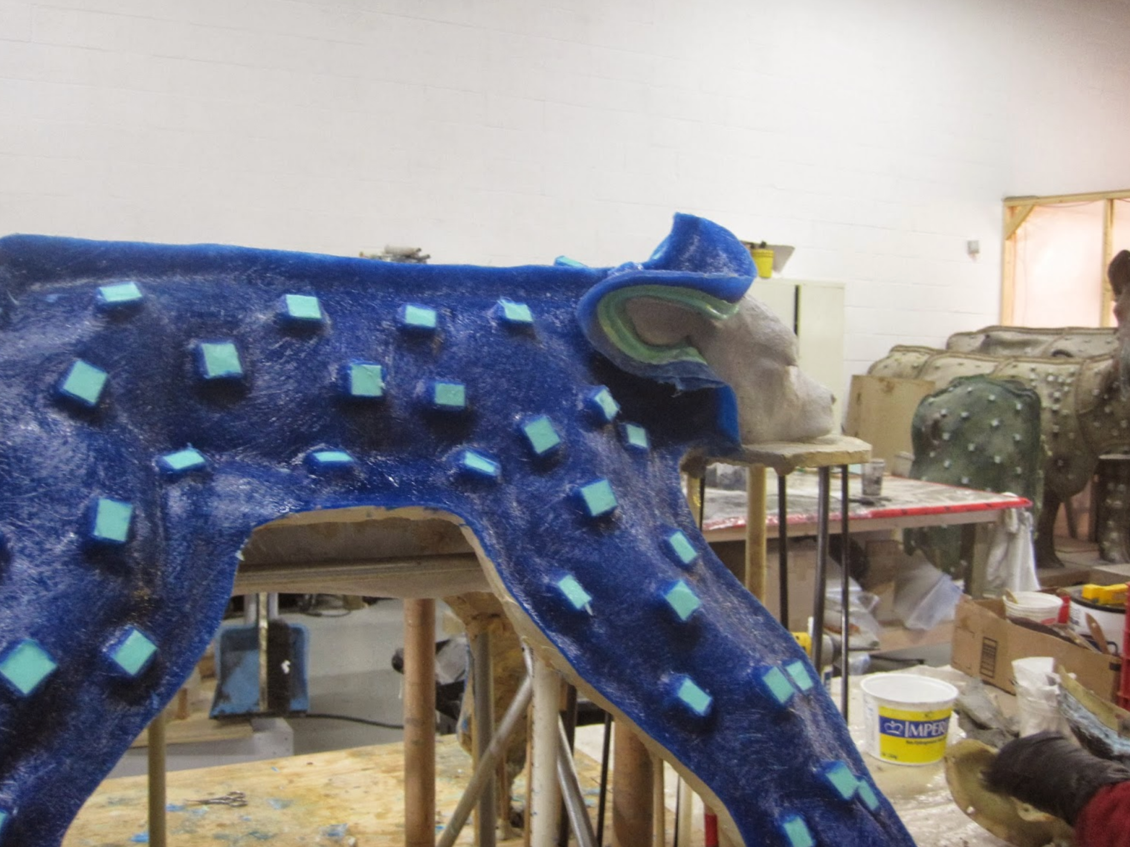
The molds have a lifespan and need to be replaced after a certain number of uses.
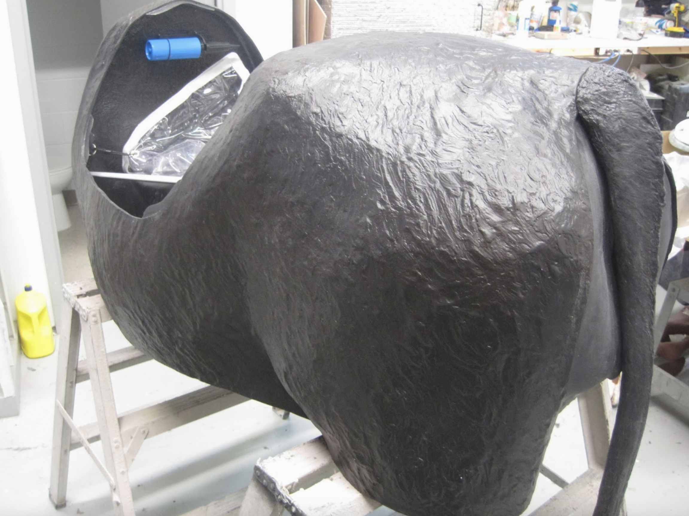
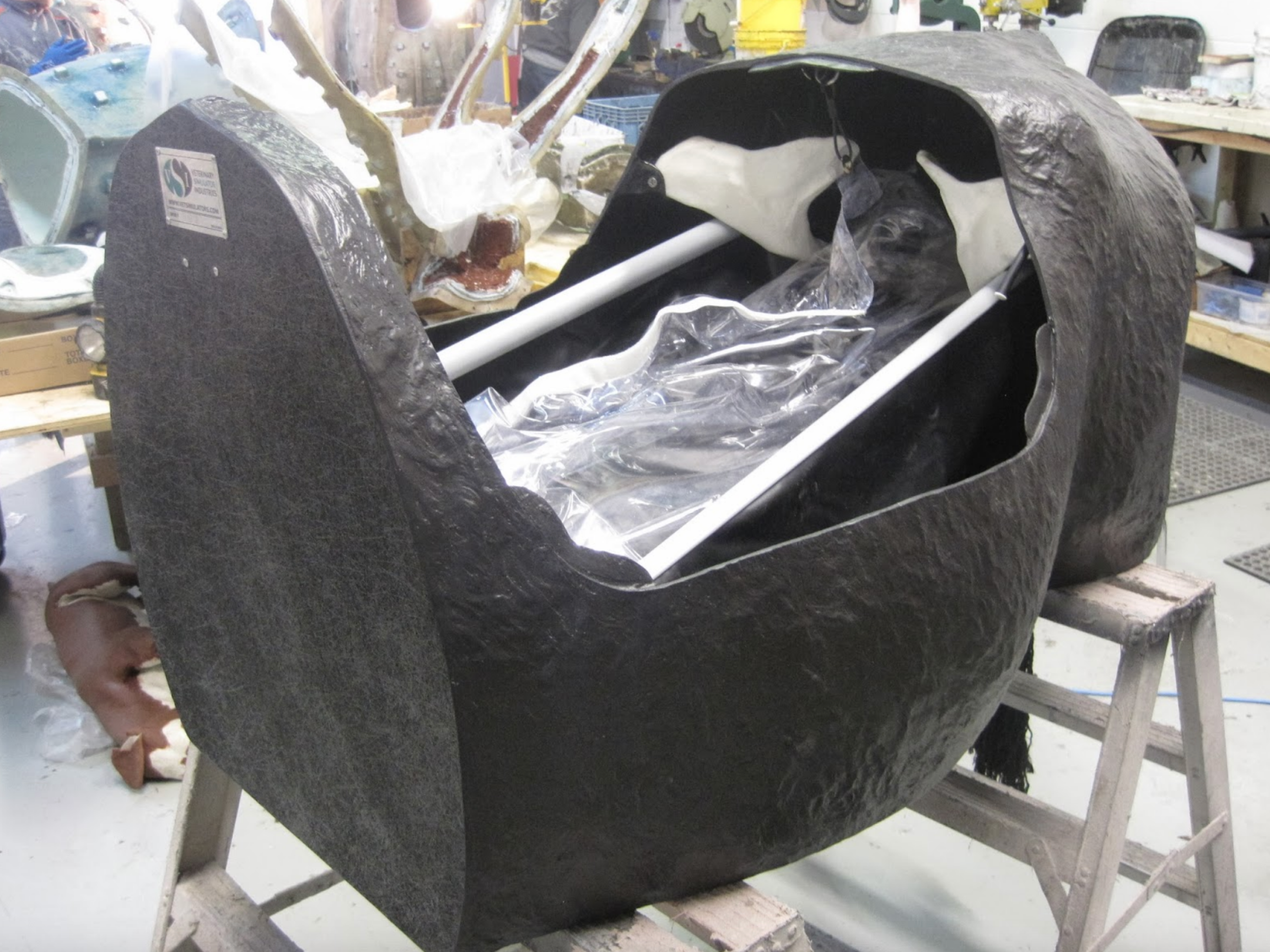
This is our compact bovine dystocia model. It has most of the functionality of our full size models except for the udder and milking capability. Because of tis compact size it reduces shipping costs and storage requirements.
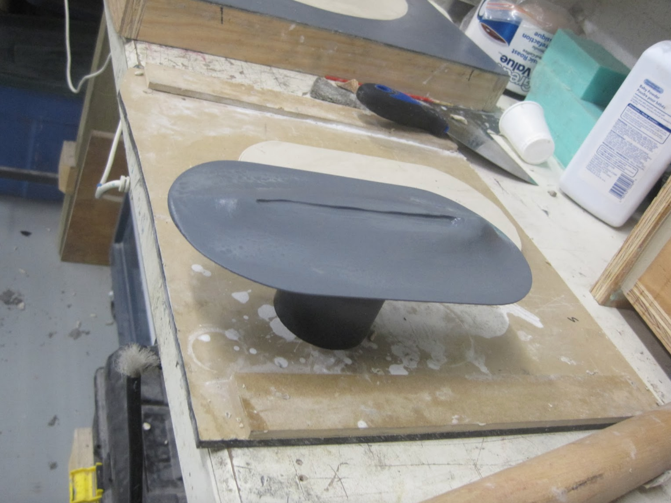
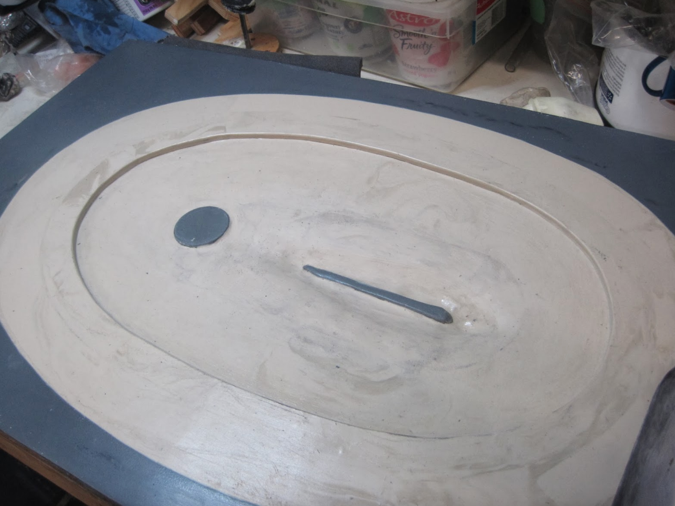
These next 3 photos show some of the stages in the development of a flexible perineum panel specifically for palpation. It will be used in conjunction with the bovine reproductive tracts


Veterinary Simulator Industries Ltd. has begun work on the second prototype for the equine neck/jugular venipuncture/intramuscular injection model. We hope to have a functioning model ready by the 2015 INVEST conference in Hanover, Germany on September 14-16th www.tiho-hannover.de/studium-lehre/…/invest-2015/.
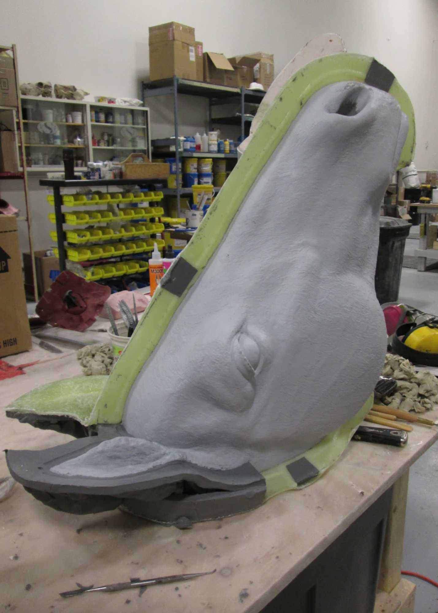
These photos show the original model which has been separated into its primary components, head, neck, shoulders. These pieces are in the process of being molded.
Once these molds are completed various materials will be then evaluated using the molds. Hopefully, if the prototypes function properly and no major changes are required, these molds will also serve as production molds.
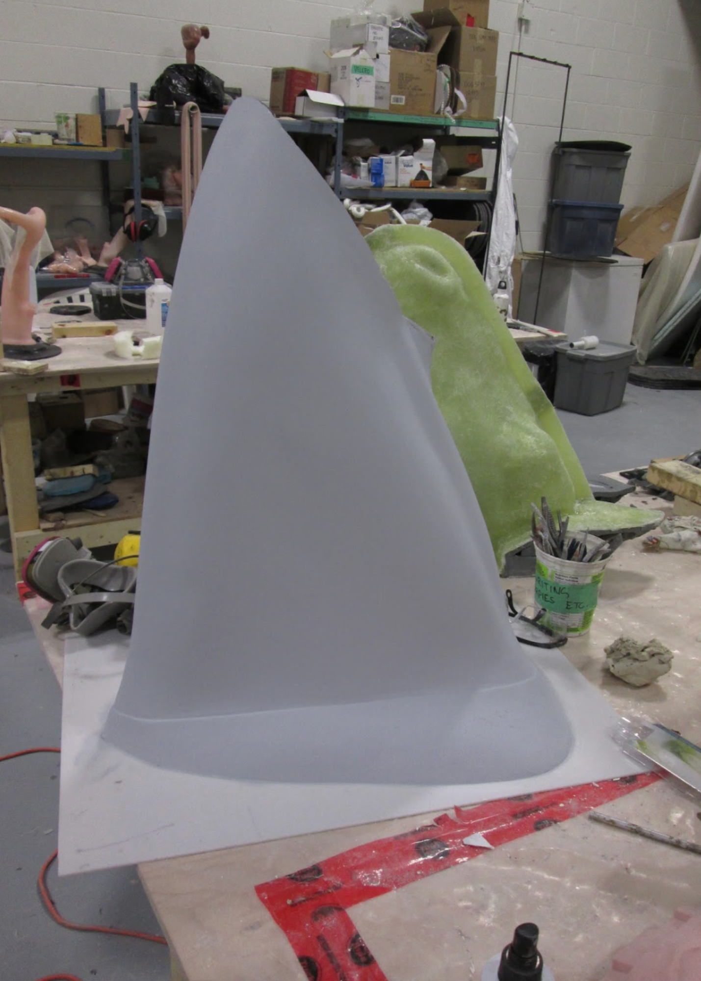
The model will be comprised of three main components, head neck, and shoulders that will fit onto an adjustable stand. The neck portion will house the venipuncture components and will have a replaceable skin of a material yet to be determined, along with an easily replaceable simulated jugular vein and intramuscular injection pad.
We are hoping a type of laminated fabric can be utilized for the skin as it could be punctured hundreds of times without damage and is relatively inexpensive.
In future we plan on adding more functions to the model such as eye medicine, facial nerves, twitch and bit/bridle.
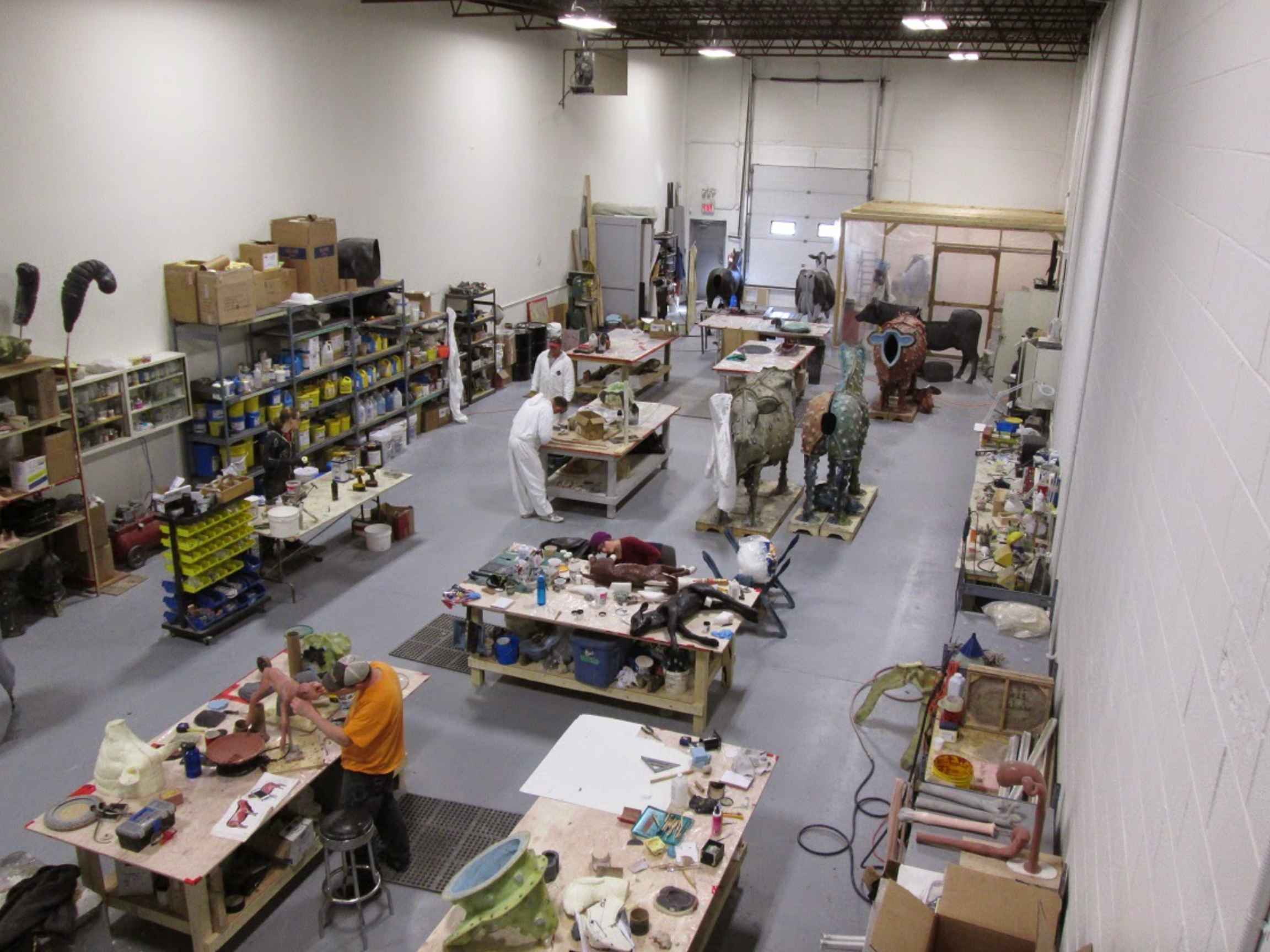
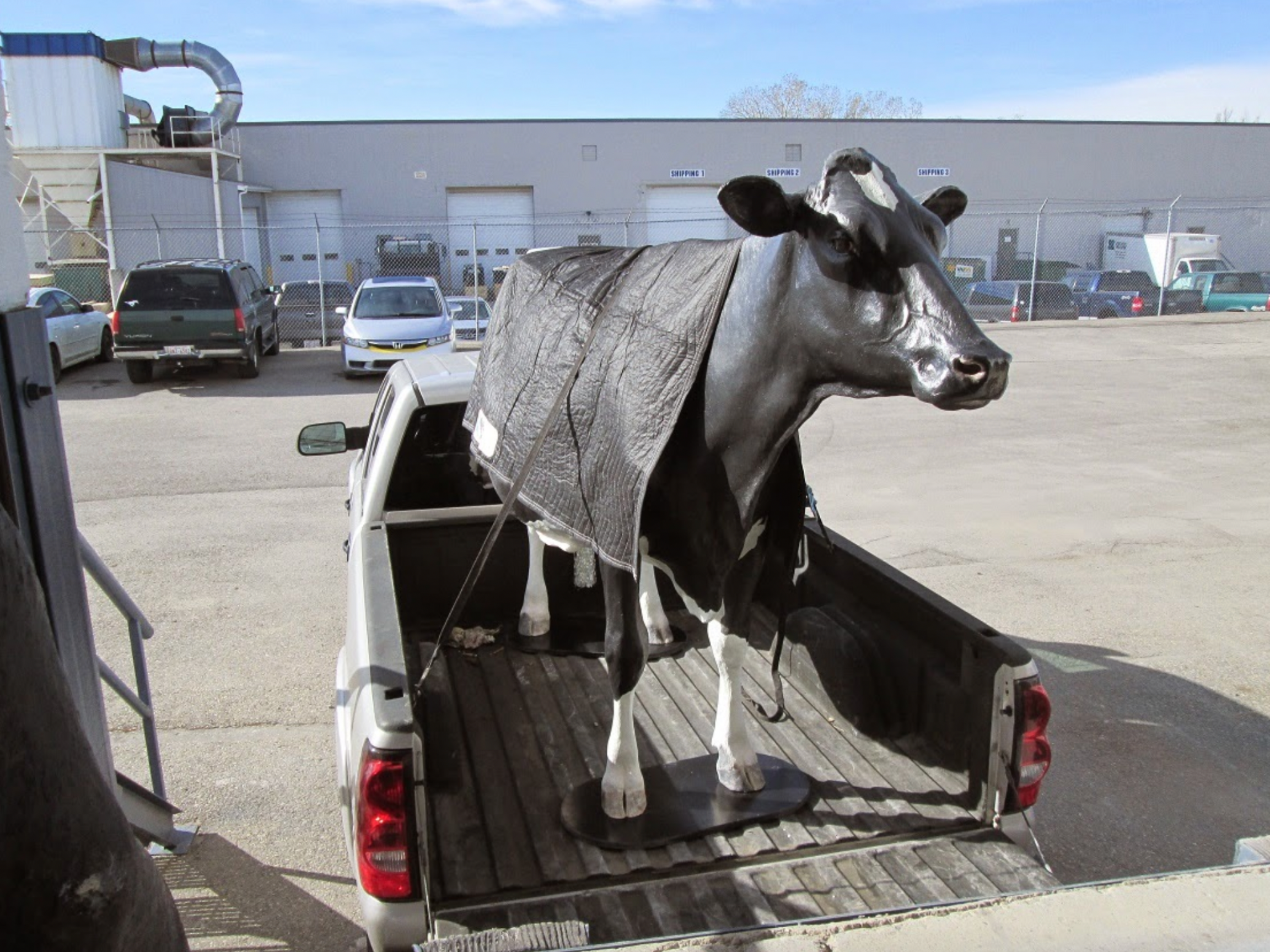
his portrait Holstein dystocia model is loaded up and heading over for crating in preparation for her trip to Iowa State University.
Her paint pattern is designed to match a Holstein named “Frosty” , a famous cow owned by an Iowa State alumni.
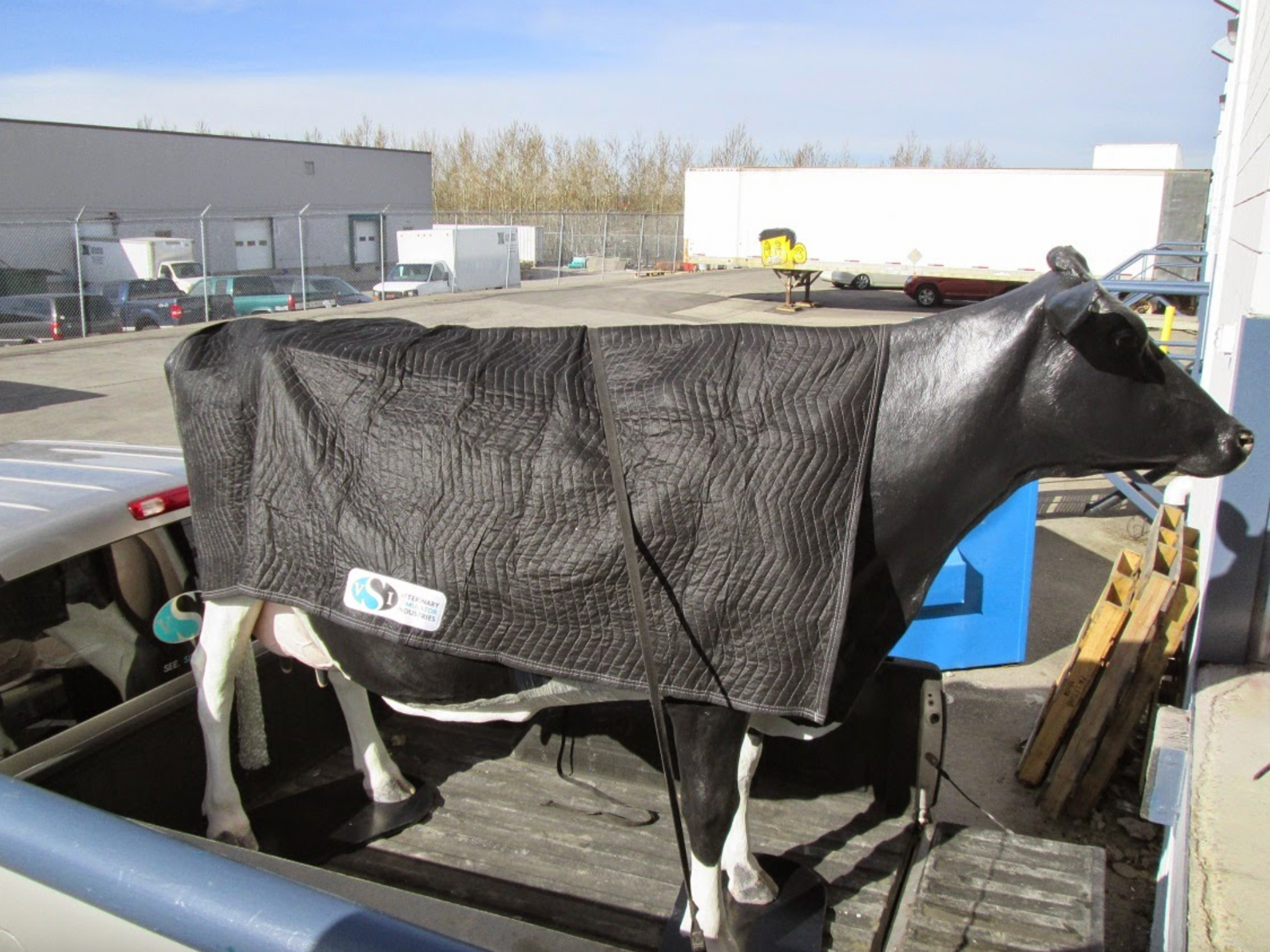
From crating she will travel by truck to Ames , Iowa and hopefully arrive within the next couple of weeks where she will be received by
Patrick G. Halbur DVM, MS, PhD
Professor and Chair, Department of Veterinary Diagnostic and Production Animal Medicine, and Dr. Pat Philips. at the College of Veterinary Medicine
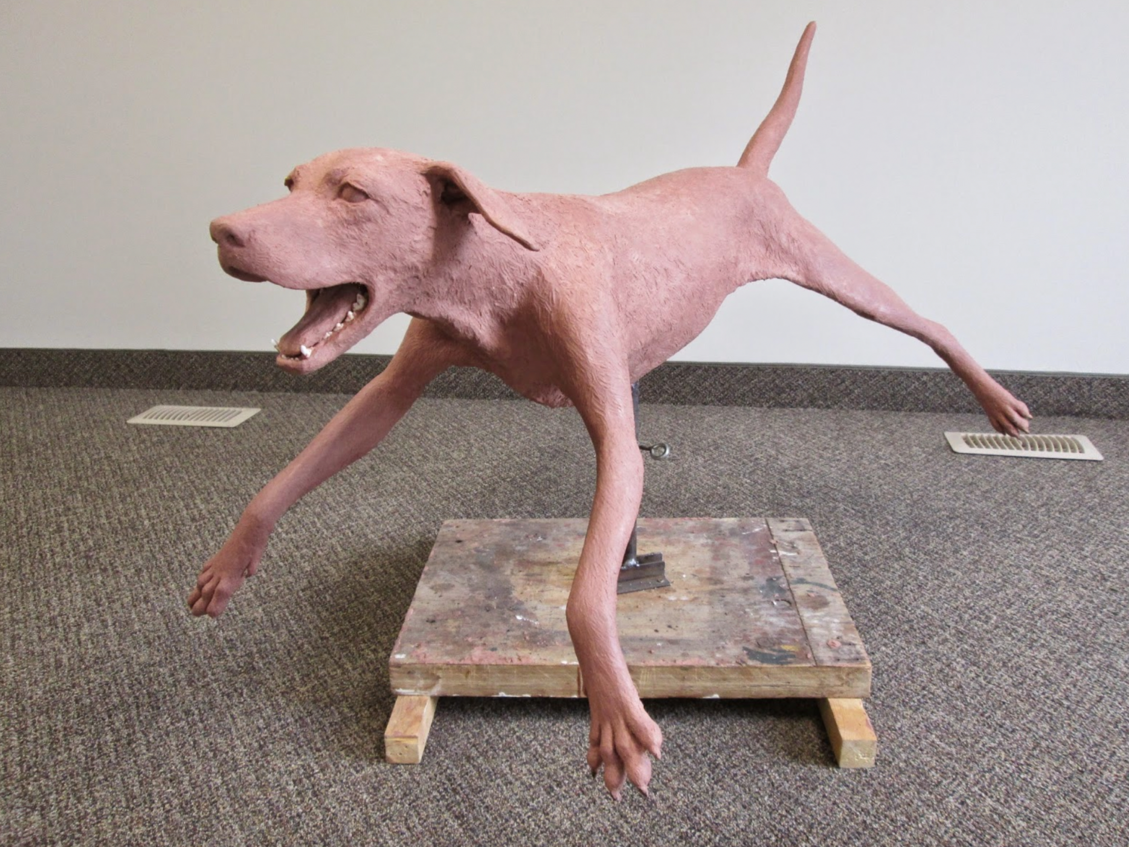
These next images show the original sculpture of our future canine simulator. It has been sculpted on an actual canine skeleton to ensure accuracy of the anatomy, positioning of joints, and allows us to cast the bones for a radiology limb model in future.
The intent of the sculpture was to be a mid sized and somewhat “generic” canine breed, with a normal chest shape.

Some of the functionality we would like to incorporate are:
Various intubation techniques
male and female catheterization bandaging limb(s)
limb venipuncture thoracentesis radiology limb(s) restraint training epidural incorporate spay/neuter function ear exams teeth mouths exams.
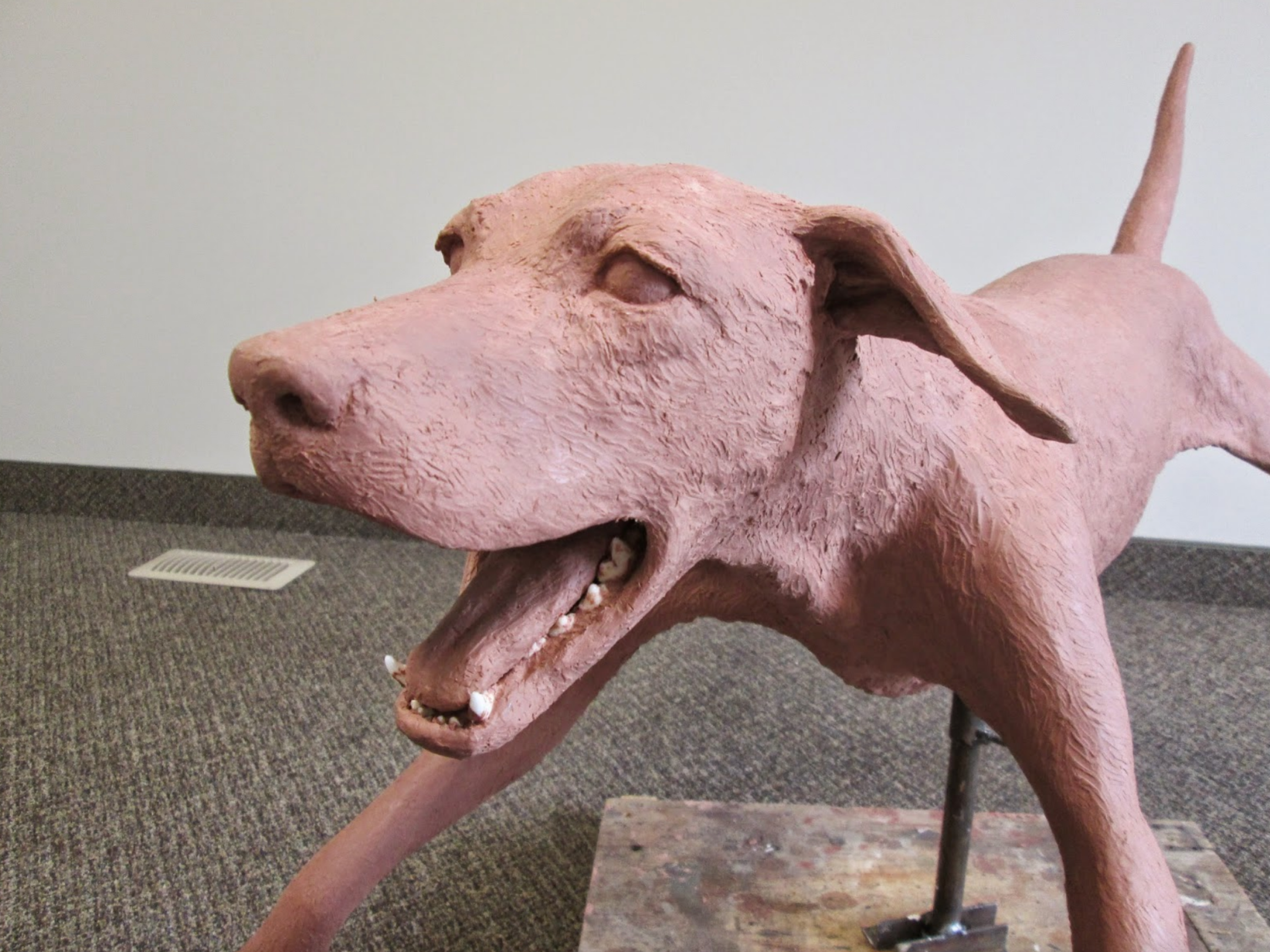
The actual canine skull and lower jaw were incorporated into the sculpture as well once again for accuracy.

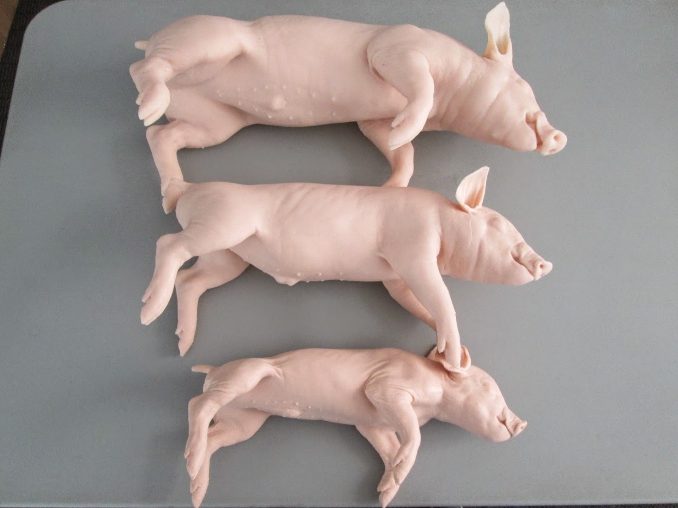
This image shows our “three little pigs” They are modeled from actual neonate piglets in three different sizes, 2-3 kg, 5-6 kg, and 7-8 kg. The piglets are for training non-penetrating captive bolt euthanasia. They are rubber with a foam core to make them accurate weights for the size.
We are in the process of modifying them so that the heads are removable and replaceable when they become damaged during training.
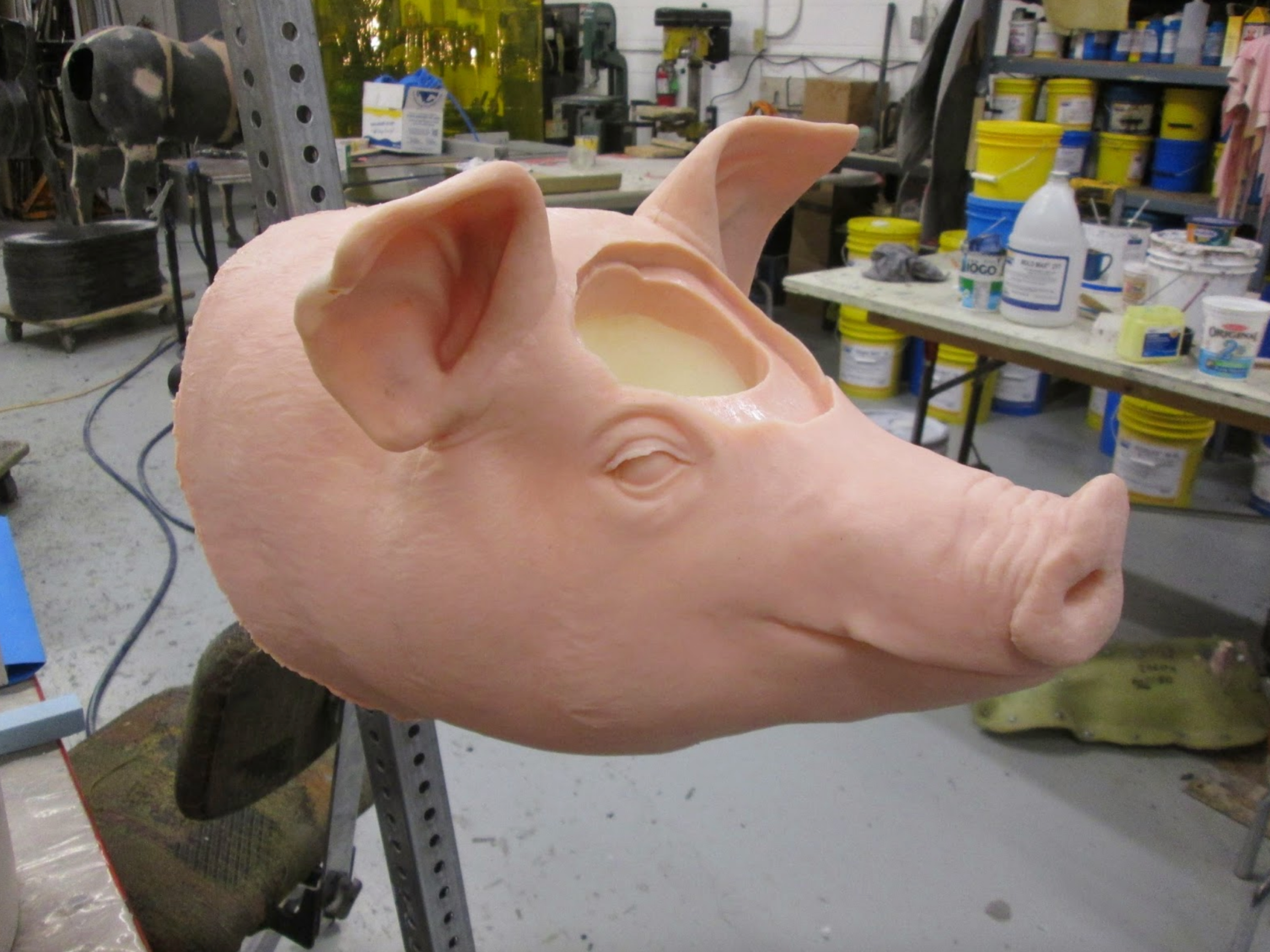
As part of our penetrating captive bolt training models we also have this market hog model.
The larger models have a removable canister comprised of a skin patch,skull plate, brain case, and brain. The head is fitted onto a stand that is adjustable for height, head tilt and head rotation.
Once targeted and shot the canister can be opened and the bolt trajectory and depth can be viewed.
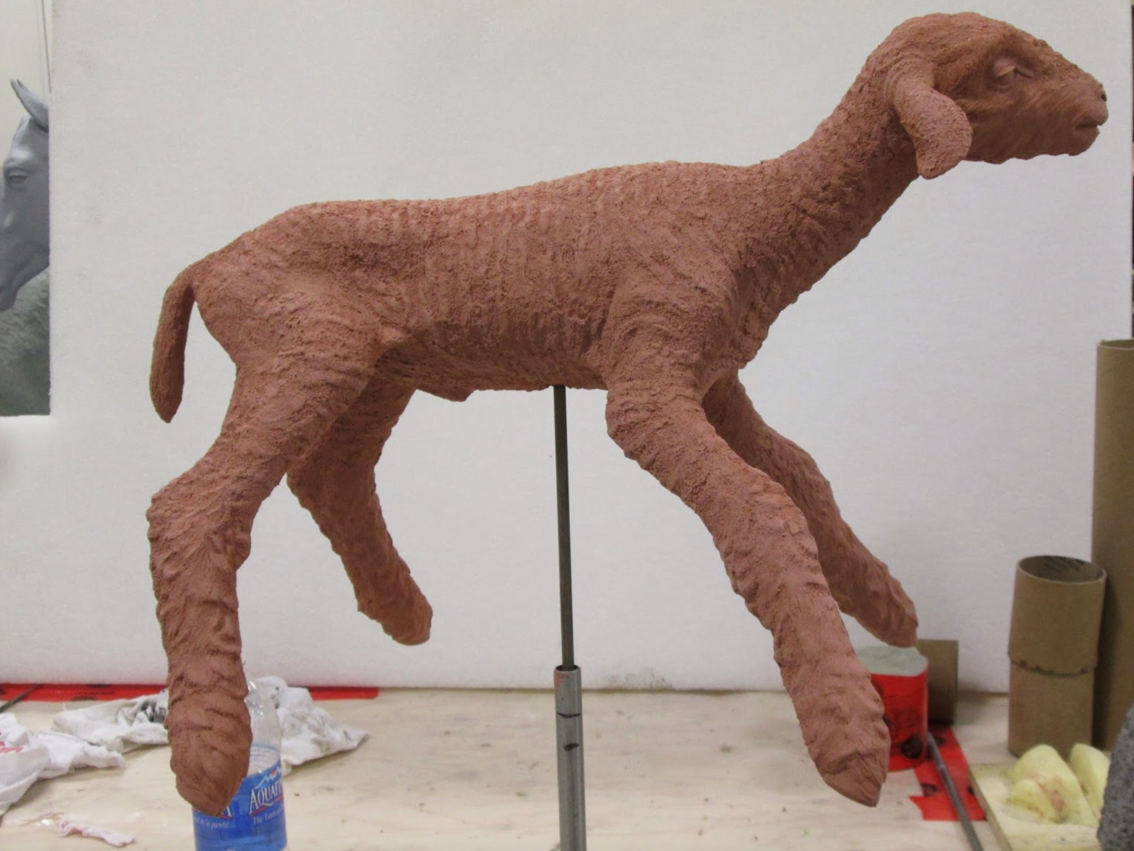
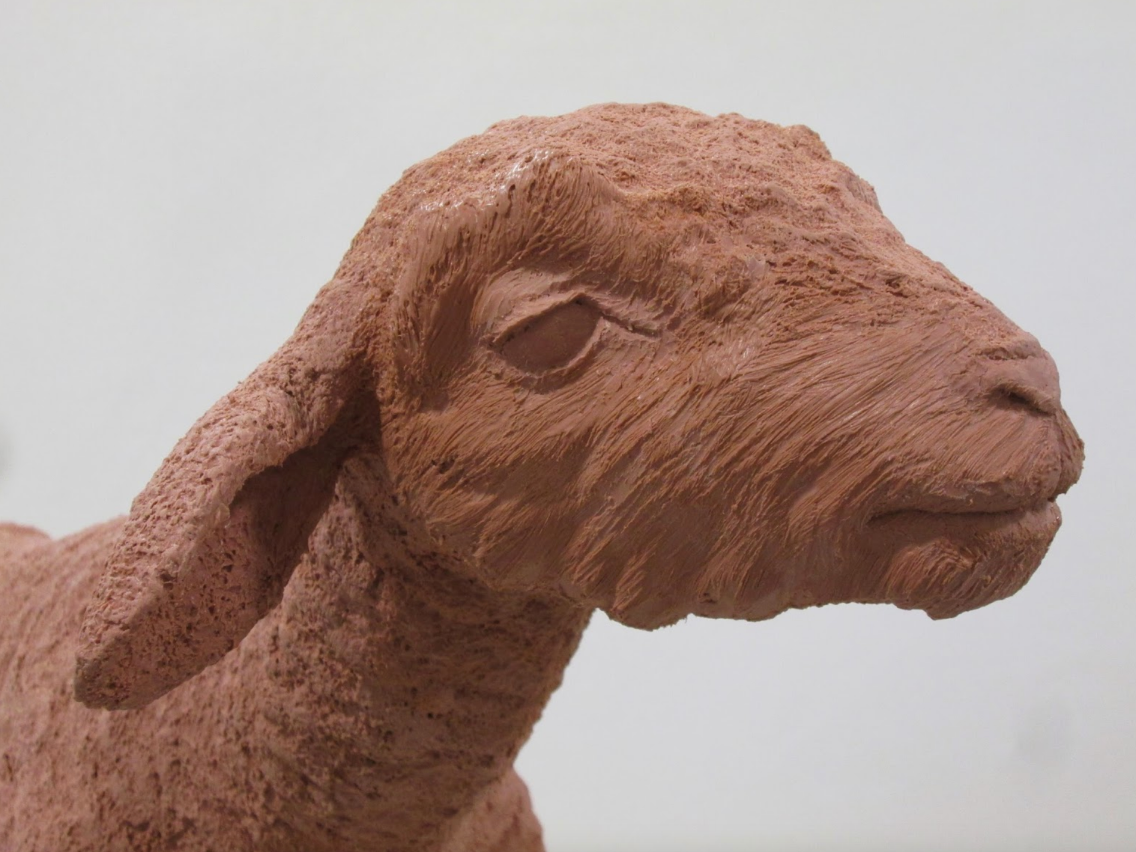
Pictured here is the 2 week old neonate lamb sculpture which will also become part of our captive bolt training system.
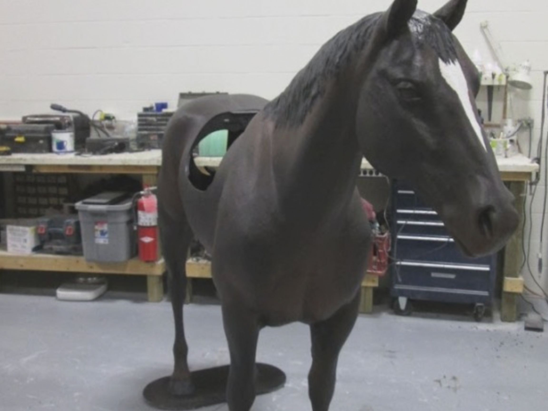
This equine colic/palpation model is ready for final assembly before being crated and shipped to Virginia-Maryland College of Veterinary Medicine.
The equine models now have a palpable aorta included, along with a portion of body wall to aid in instructing students with equine palpation.
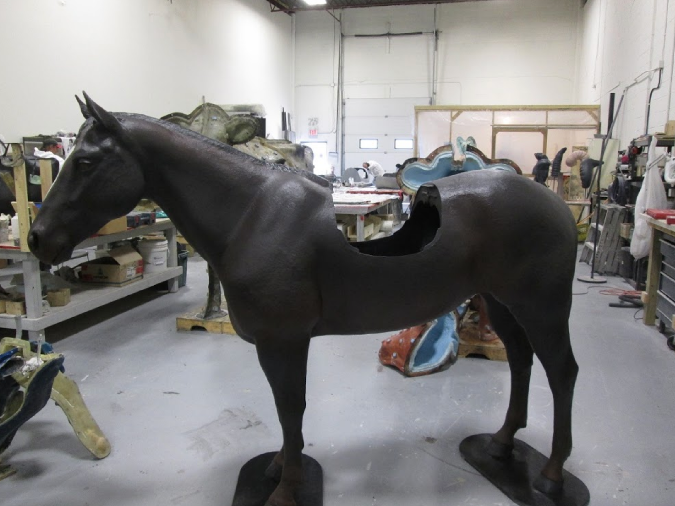
We will be shipping out models to Antwerp, Belgium and University of Tennessee in the near future.
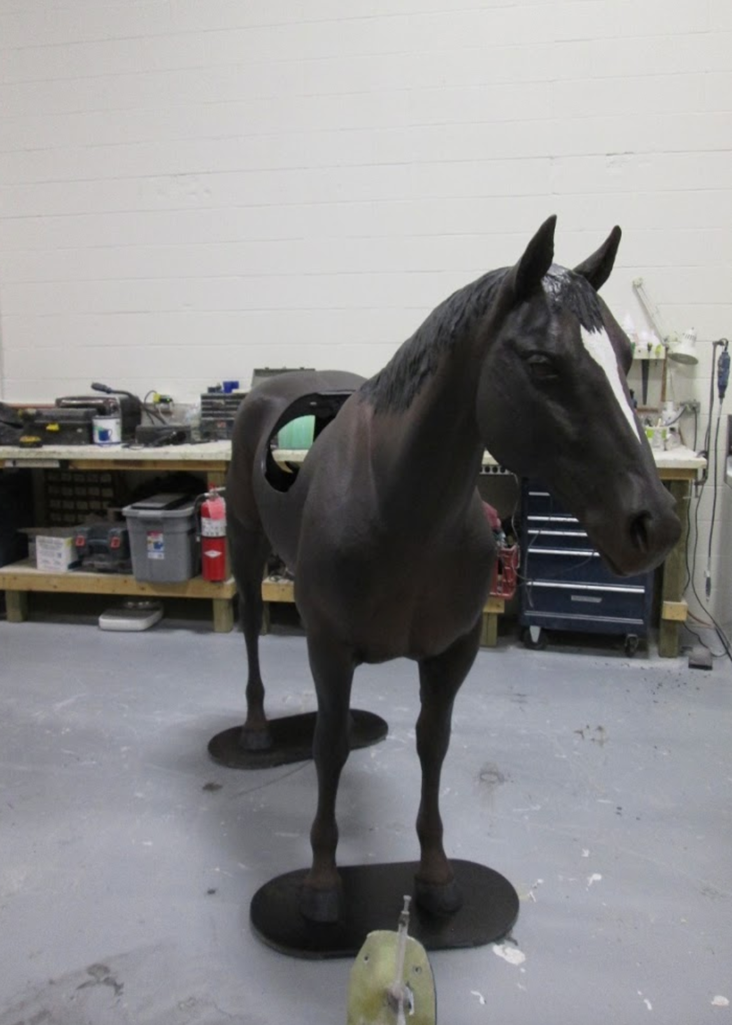
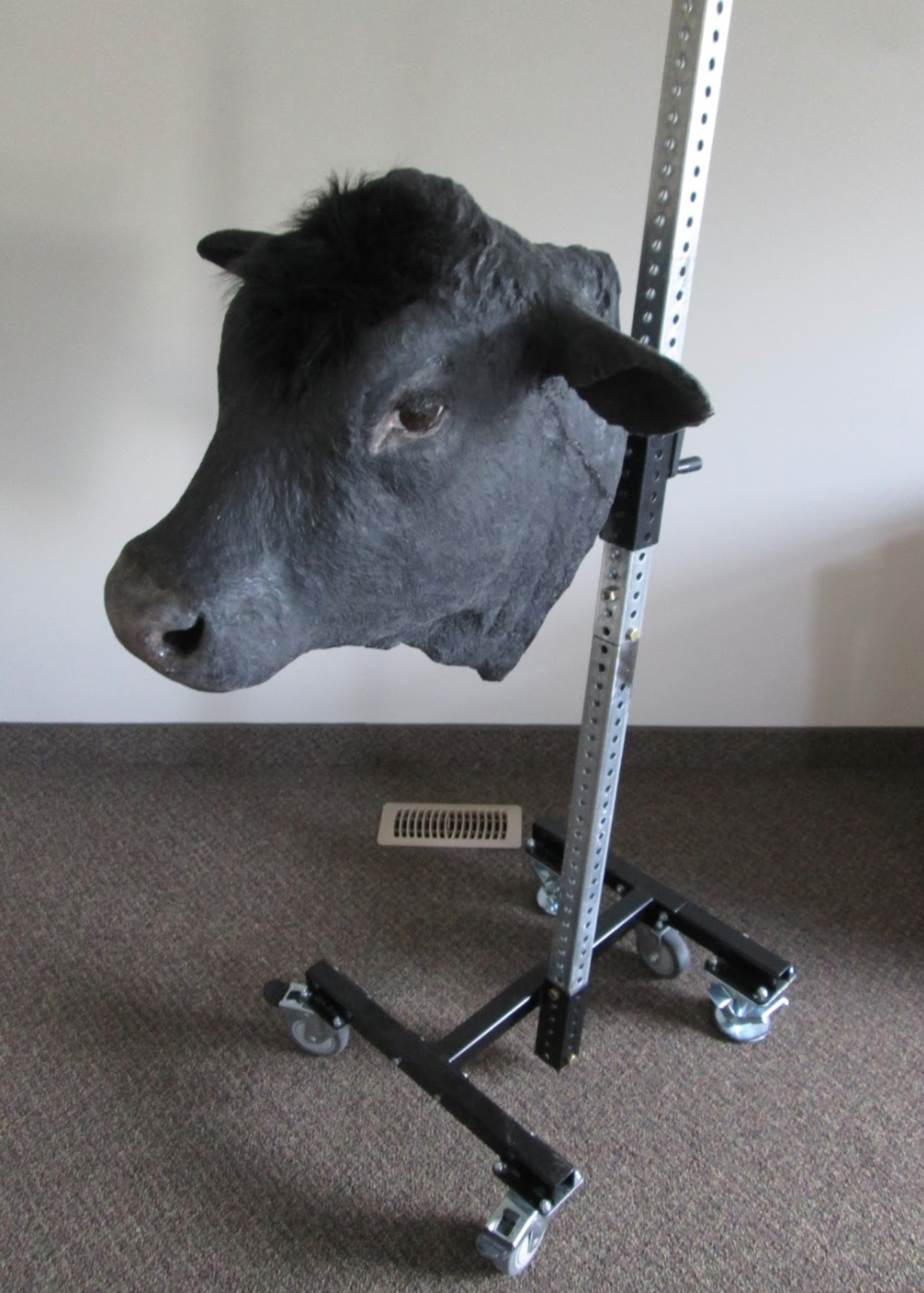
Veterinary Simulator Industries Ltd has been working on a captive bolt bovine stunning simulator with funding from the Alberta Livestock and Meat Agency.
This prototype model is based on a market steer and utilizes a replaceable brain canister system.
The canister (shown below) has representations of the skin, skull and brain.
This example also has a three dimensional target area included that represents the optimal placement for an effective stun and could be used for operator competency certification.
We have have also created canisters that would only be used for targeting training, and for getting experience in firing the captive bolt gun.
Now that the prototype has been completed it will be further evaluated at Olds Agricultural College in the National Meat Training Center.
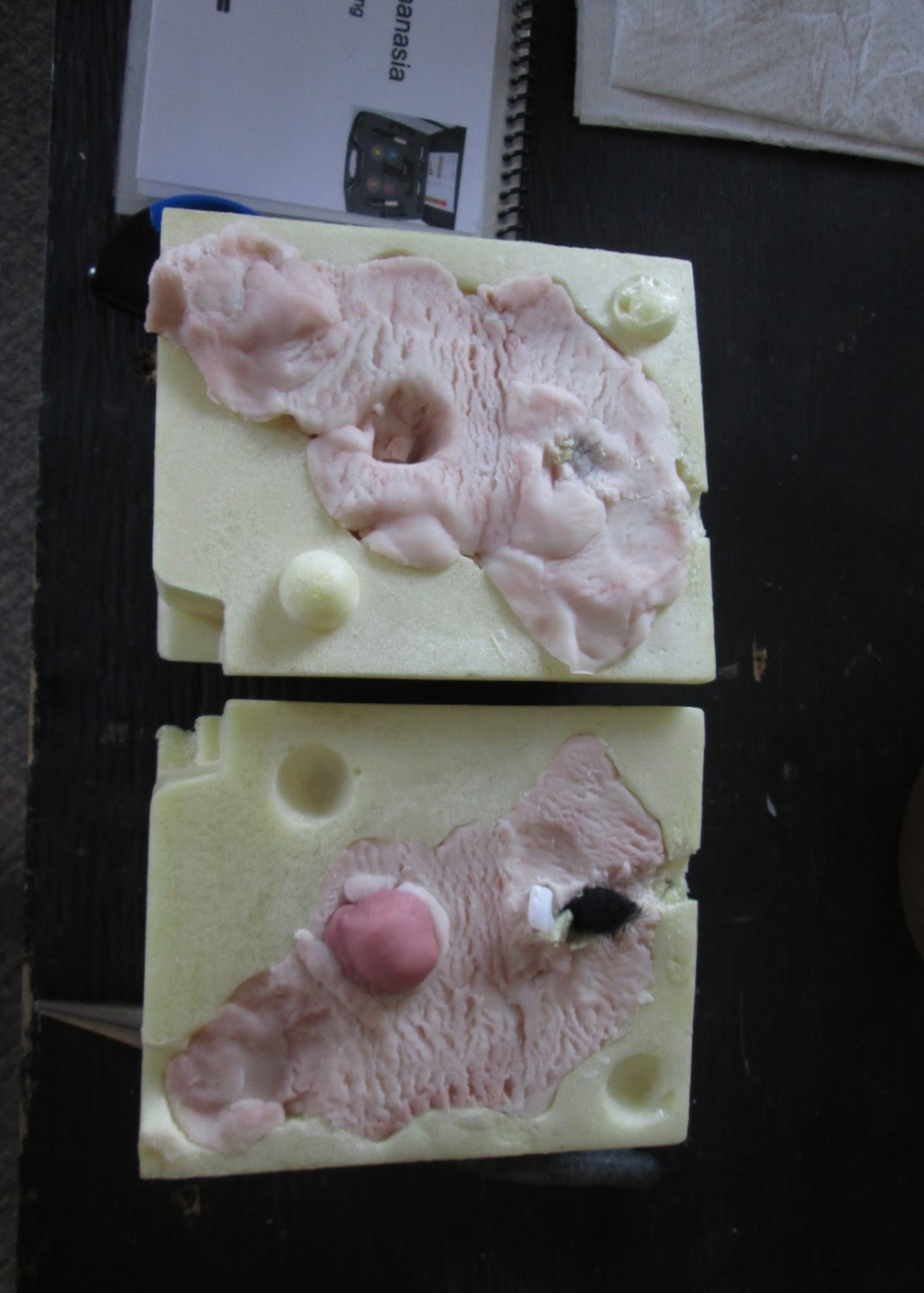
This opened canister shows the path left by a captive bolt shot along with a piece of skin and skull at the end of the bolt’s travel.
Dr. Bob Hollowaychuk, as part of the original ALMA proposal, test shot this model with a CASH model captive bolt.
Dr. Holowaychuk whom recently retired following a successful thirty-five year career in regulatory inspection services at both the federal and provincial level will review the simulation model and accompanying training materials to ensure that the system meets all federal and provincial regulations.


